4K28

2.15 Angstrom resolution crystal structure of a shikimate dehydrogenase family protein from Pseudomonas putida KT2440 in complex with NAD+


Experimental Data Snapshot

  • Method: X-RAY DIFFRACTION
  • Resolution: 2.15 Å
  • R-Value Free: 0.238 
  • R-Value Work: 0.179 
  • R-Value Observed: 0.182 

wwPDB Validation   3D Report Full Report


Ligand Structure Quality Assessment 


This is version 1.1 of the entry. See complete history


Literature

Insights into the function of RifI2: structural and biochemical investigation of a new shikimate dehydrogenase family protein.

Peek, J.Garcia, C.Lee, J.Christendat, D.

(2013) Biochim Biophys Acta 1834: 516-523

  • DOI: https://doi.org/10.1016/j.bbapap.2012.10.016
  • Primary Citation of Related Structures:  
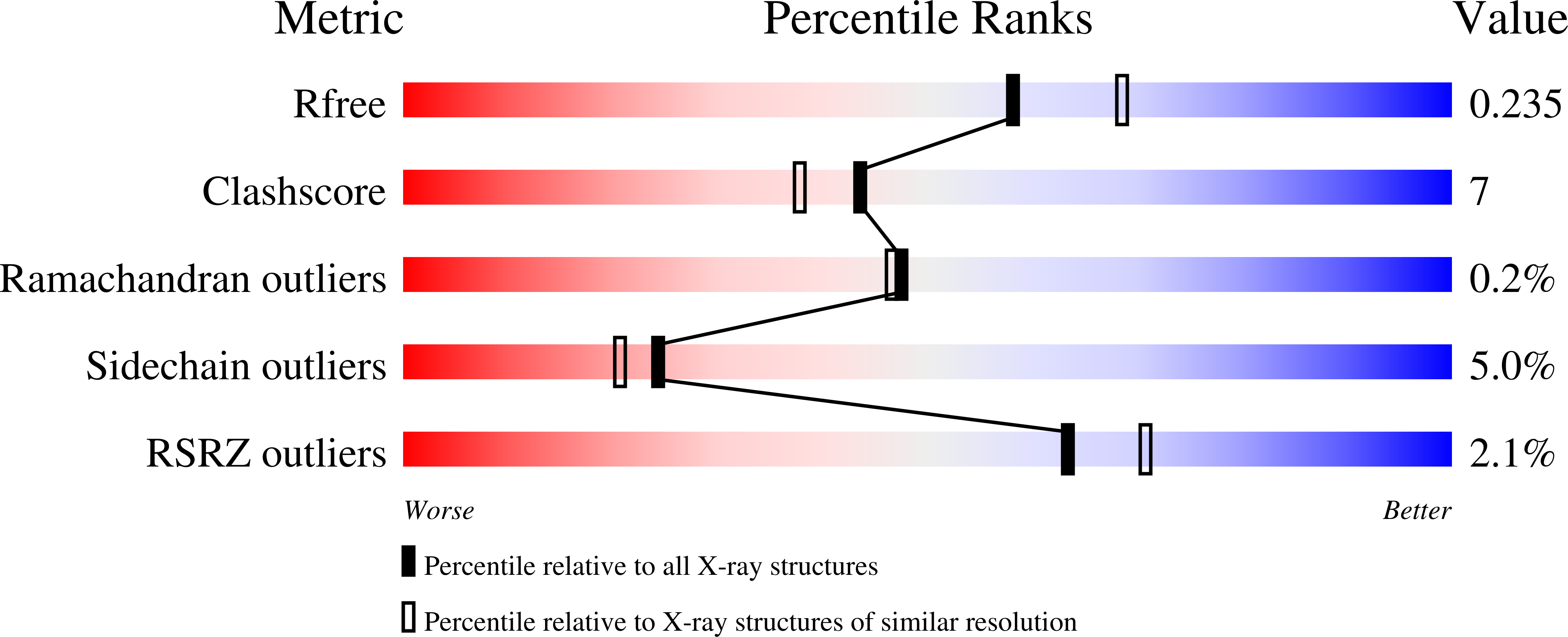
    4K28

  • PubMed Abstract: 

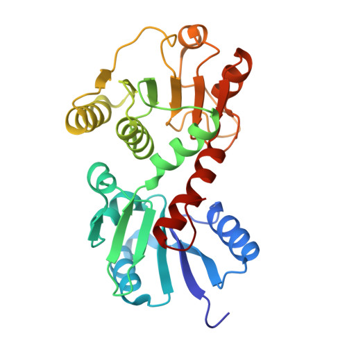
    The shikimate dehydrogenase (SDH) family consists of enzymes with diverse roles in secondary metabolism. The two most widespread members of the family, AroE and YdiB, function in amino acid biosynthesis and quinate catabolism, respectively. Here, we have determined the crystal structure of an SDH homolog belonging to the RifI class, a group of enzymes with proposed roles in antibiotic biosynthesis. The structure of RifI2 from Pseudomonas putida exhibits a number of distinctive features, including a substantial C-terminal truncation and an atypical mode of oligomerization. The active site of the enzyme contains substrate- and cofactor-binding motifs that are significantly different from those of any previously characterized member of the SDH family. These features are reflected in the novel kinetic properties of the enzyme. RifI2 exhibits much lower activity using shikimate as a substrate than AroE, and a strong preference for NAD(+) instead of NADP(+) as a cofactor. Moreover, the enzyme has only trace activity using quinate, unlike YdiB. Cocrystallization of RifI2 with NAD(+) provided the opportunity to determine the mode of cofactor selectivity employed by the enzyme. We complemented this analysis by probing the role of a strictly conserved residue in the cofactor-binding domain, Asn193, by site directed mutagenesis. This study presents the first crystal structure and formal kinetic characterization of a new NAD(+)-dependent member of the SDH family.


  • Organizational Affiliation

    Department of Cell and Systems Biology, University of Toronto, Toronto, Ontario, Canada M5S 3B2.


Macromolecules
Find similar proteins by:  (by identity cutoff)  |  3D Structure
Entity ID: 1
MoleculeChains Sequence LengthOrganismDetailsImage
Shikimate dehydrogenase family protein
A, B
269Pseudomonas putida KT2440Mutation(s): 0 
Gene Names: pp2608PP_2608
UniProt
Find proteins for Q88JP1 (Pseudomonas putida (strain ATCC 47054 / DSM 6125 / CFBP 8728 / NCIMB 11950 / KT2440))
Explore Q88JP1 
Go to UniProtKB:  Q88JP1
Entity Groups  
Sequence Clusters30% Identity50% Identity70% Identity90% Identity95% Identity100% Identity
UniProt GroupQ88JP1
Sequence Annotations
Expand
  • Reference Sequence
Small Molecules
Ligands 3 Unique
IDChains Name / Formula / InChI Key2D Diagram3D Interactions
NAD
Query on NAD

Download Ideal Coordinates CCD File 
C [auth A],
G [auth B]
NICOTINAMIDE-ADENINE-DINUCLEOTIDE
C21 H27 N7 O14 P2
BAWFJGJZGIEFAR-NNYOXOHSSA-N
MN
Query on MN

Download Ideal Coordinates CCD File 
D [auth A]
E [auth A]
F [auth A]
I [auth B]
J [auth B]
D [auth A],
E [auth A],
F [auth A],
I [auth B],
J [auth B],
K [auth B]
MANGANESE (II) ION
Mn
WAEMQWOKJMHJLA-UHFFFAOYSA-N
NA
Query on NA

Download Ideal Coordinates CCD File 
H [auth B]SODIUM ION
Na
FKNQFGJONOIPTF-UHFFFAOYSA-N
Experimental Data & Validation

Experimental Data

  • Method: X-RAY DIFFRACTION
  • Resolution: 2.15 Å
  • R-Value Free: 0.238 
  • R-Value Work: 0.179 
  • R-Value Observed: 0.182 
  • Space Group: P 21 21 21
Unit Cell:
Length ( Å )Angle ( ˚ )
a = 44.437α = 90
b = 76.688β = 90
c = 153.333γ = 90
Software Package:
Software NamePurpose
CrystalCleardata collection
MOLREPphasing
PHENIXrefinement
XDSdata reduction
SCALAdata scaling

Structure Validation

View Full Validation Report


Ligand Structure Quality Assessment 


Entry History 

Deposition Data

Revision History  (Full details and data files)

  • Version 1.0: 2013-04-17
    Type: Initial release
  • Version 1.1: 2024-02-28
    Changes: Data collection, Database references, Derived calculations