4K1P

Structure of the NheA component of the Nhe toxin from Bacillus cereus


Experimental Data Snapshot

  • Method: X-RAY DIFFRACTION
  • Resolution: 2.05 Å
  • R-Value Free: 0.262 
  • R-Value Work: 0.212 
  • R-Value Observed: 0.214 

wwPDB Validation   3D Report Full Report


This is version 1.2 of the entry. See complete history


Literature

Structure of the NheA Component of the Nhe Toxin from Bacillus cereus: Implications for Function.

Ganash, M.Phung, D.Sedelnikova, S.E.Lindback, T.Granum, P.E.Artymiuk, P.J.

(2013) PLoS One 8: e74748-e74748

  • DOI: https://doi.org/10.1371/journal.pone.0074748
  • Primary Citation of Related Structures:  
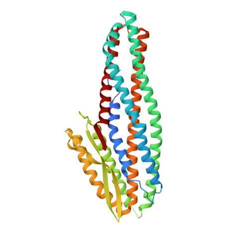
    4K1P

  • PubMed Abstract: 

    The structure of NheA, a component of the Bacillus cereus Nhe tripartite toxin, has been solved at 2.05 Å resolution using selenomethionine multiple-wavelength anomalous dispersion (MAD). The structure shows it to have a fold that is similar to the Bacillus cereus Hbl-B and E. coli ClyA toxins, and it is therefore a member of the ClyA superfamily of α-helical pore forming toxins (α-PFTs), although its head domain is significantly enlarged compared with those of ClyA or Hbl-B. The hydrophobic β-hairpin structure that is a characteristic of these toxins is replaced by an amphipathic β-hairpin connected to the main structure via a β-latch that is reminiscent of a similar structure in the β-PFT Staphylococcus aureus α-hemolysin. Taken together these results suggest that, although it is a member of an archetypal α-PFT family of toxins, NheA may be capable of forming a β rather than an α pore.


  • Organizational Affiliation

    The Krebs Institute, Department of Molecular Biology and Biotechnology, University of Sheffield, Sheffield, United Kingdom.


Macromolecules
Find similar proteins by:  (by identity cutoff)  |  3D Structure
Entity ID: 1
MoleculeChains Sequence LengthOrganismDetailsImage
NheA
A, B, C, D, E
A, B, C, D, E, F, G, H
360Bacillus cereusMutation(s): 0 
Gene Names: nheAnheABC (operon)
UniProt
Find proteins for Q3Y6N6 (Bacillus cereus)
Explore Q3Y6N6 
Go to UniProtKB:  Q3Y6N6
Entity Groups  
Sequence Clusters30% Identity50% Identity70% Identity90% Identity95% Identity100% Identity
UniProt GroupQ3Y6N6
Sequence Annotations
Expand
  • Reference Sequence
Experimental Data & Validation

Experimental Data

  • Method: X-RAY DIFFRACTION
  • Resolution: 2.05 Å
  • R-Value Free: 0.262 
  • R-Value Work: 0.212 
  • R-Value Observed: 0.214 
  • Space Group: C 1 2 1
Unit Cell:
Length ( Å )Angle ( ˚ )
a = 309.008α = 90
b = 58.242β = 110.61
c = 172.999γ = 90
Software Package:
Software NamePurpose
REFMACrefinement
PDB_EXTRACTdata extraction
ADSCdata collection
xia2data reduction
SCALEPACKdata scaling
SHELXSphasing

Structure Validation

View Full Validation Report


Entry History 

Deposition Data

Revision History  (Full details and data files)

  • Version 1.0: 2013-09-18
    Type: Initial release
  • Version 1.1: 2013-10-02
    Changes: Database references
  • Version 1.2: 2024-02-28
    Changes: Data collection, Database references, Derived calculations