4K0U

Pilotin/secretin peptide Complex


Experimental Data Snapshot

  • Method: X-RAY DIFFRACTION
  • Resolution: 2.15 Å
  • R-Value Free: 0.276 
  • R-Value Work: 0.210 
  • R-Value Observed: 0.213 

wwPDB Validation   3D Report Full Report


This is version 1.2 of the entry. See complete history


Literature

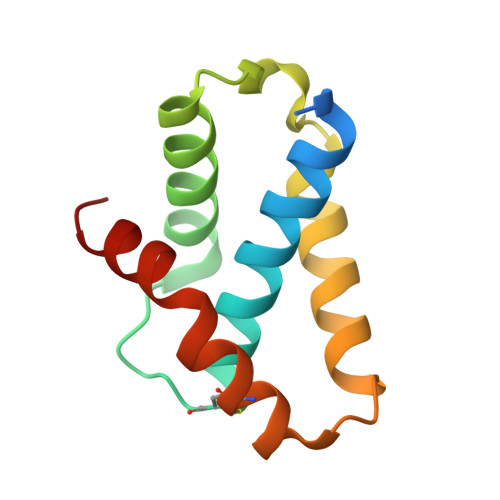
Anatomy of secretin binding to the Dickeya dadantii type II secretion system pilotin.

Rehman, S.Gu, S.Shevchik, V.E.Pickersgill, R.W.

(2013) Acta Crystallogr D Biol Crystallogr 69: 1381-1386

  • DOI: https://doi.org/10.1107/S0907444913007658
  • Primary Citation of Related Structures:  
    4K0U

  • PubMed Abstract: 

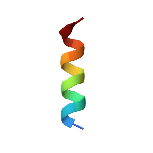
    The secretins are a family of large multimeric channels in the outer membrane of Gram-negative bacteria that are involved in protein export. In Dickeya dadantii and many other pathogenic bacteria, the lipoprotein pilotin targets the secretin subunits to the outer membrane, allowing a functional type II secretion system to be assembled. Here, the crystal structure of the C-terminal peptide of the secretin subunit bound to its cognate pilotin is reported. In solution, this C-terminal region of the secretin is nonstructured. The secretin peptide folds on binding to the pilotin to form just under four turns of α-helix which bind tightly up against the first helix of the pilotin so that the hydrophobic residues of the secretin helix can bind to the hydrophobic surface of the pilotin. The secretin helix binds parallel to the first part of the fourth helix of the pilotin. An N-capping aspartate encourages helix formation and binding by interacting favourably with the helix dipole of the helical secretin peptide. The structure of the secretin-pilotin complex of the phytopathogenic D. dadantii described here is a paradigm for this interaction in the OutS-PulS family of pilotins, which is essential for the correct assembly of the type II secretion system of several potent human adversaries, including enterohaemorrhagic Escherichia coli and Klebsiella oxytoca.


  • Organizational Affiliation

    School of Biological and Chemical Sciences, Queen Mary University of London, Mile End Road, London E1 4NS, England.


Macromolecules
Find similar proteins by:  (by identity cutoff)  |  3D Structure
Entity ID: 1
MoleculeChains Sequence LengthOrganismDetailsImage
Lipoprotein OutS106Dickeya dadantii 3937Mutation(s): 0 
Gene Names: Dda3937_02411outSOutS Dda3937_02411
UniProt
Find proteins for Q01567 (Dickeya dadantii (strain 3937))
Explore Q01567 
Go to UniProtKB:  Q01567
Entity Groups  
Sequence Clusters30% Identity50% Identity70% Identity90% Identity95% Identity100% Identity
UniProt GroupQ01567
Sequence Annotations
Expand
  • Reference Sequence

Find similar proteins by:  Sequence   |   3D Structure  

Entity ID: 2
MoleculeChains Sequence LengthOrganismDetailsImage
Type II secretion system protein D15Dickeya dadantii 3937Mutation(s): 0 
UniProt
Find proteins for Q01565 (Dickeya dadantii (strain 3937))
Explore Q01565 
Go to UniProtKB:  Q01565
Entity Groups  
Sequence Clusters30% Identity50% Identity70% Identity90% Identity95% Identity100% Identity
UniProt GroupQ01565
Sequence Annotations
Expand
  • Reference Sequence
Experimental Data & Validation

Experimental Data

  • Method: X-RAY DIFFRACTION
  • Resolution: 2.15 Å
  • R-Value Free: 0.276 
  • R-Value Work: 0.210 
  • R-Value Observed: 0.213 
  • Space Group: P 65 2 2
Unit Cell:
Length ( Å )Angle ( ˚ )
a = 53.602α = 90
b = 53.602β = 90
c = 142.441γ = 120
Software Package:
Software NamePurpose
MxCuBEdata collection
MOLREPphasing
REFMACrefinement
XDSdata reduction
SCALAdata scaling

Structure Validation

View Full Validation Report


Entry History 

Deposition Data

Revision History  (Full details and data files)

  • Version 1.0: 2013-05-15
    Type: Initial release
  • Version 1.1: 2015-06-24
    Changes: Database references
  • Version 1.2: 2023-11-08
    Changes: Data collection, Database references, Refinement description