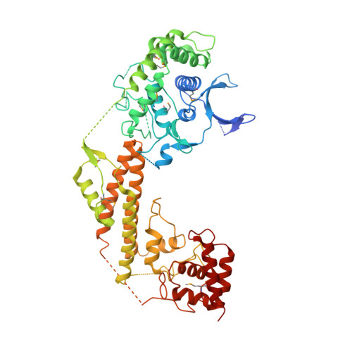
Structural analyses of Legionella LepB reveal a new GAP fold that catalytically mimics eukaryotic RasGAP
Yu, Q., Hu, L., Yao, Q., Zhu, Y., Dong, N., Wang, D.-C., Shao, F.(2013) Cell Res 23: 775-787
- PubMed: 23588383
- DOI: https://doi.org/10.1038/cr.2013.54
- Primary Citation of Related Structures:
4JVS, 4JW1 - PubMed Abstract:
Rab GTPases are emerging targets of diverse bacterial pathogens. Here, we perform biochemical and structural analyses of LepB, a Rab GTPase-activating protein (GAP) effector from Legionella pneumophila. We map LepB GAP domain to residues 313-618 and show that the GAP domain is Rab1 specific with a catalytic activity higher than the canonical eukaryotic TBC GAP and the newly identified VirA/EspG family of bacterial RabGAP effectors. Exhaustive mutation analyses identify Arg444 as the arginine finger, but no catalytically essential glutamine residues. Crystal structures of LepB313-618 alone and the GAP domain of Legionella drancourtii LepB in complex with Rab1-GDP-AlF3 support the catalytic role of Arg444, and also further reveal a 3D architecture and a GTPase-binding mode distinct from all known GAPs. Glu449, structurally equivalent to TBC RabGAP glutamine finger in apo-LepB, undergoes a drastic movement upon Rab1 binding, which induces Rab1 Gln70 side-chain flipping towards GDP-AlF3 through a strong ionic interaction. This conformationally rearranged Gln70 acts as the catalytic cis-glutamine, therefore uncovering an unexpected RasGAP-like catalytic mechanism for LepB. Our studies highlight an extraordinary structural and catalytic diversity of RabGAPs, particularly those from bacterial pathogens.
Organizational Affiliation:
National Laboratory of Biomacromolecules, Institute of Biophysics, Chinese Academy of Sciences, Beijing 100101, China.