4JMH

Crystal structure of synthetic protein in complex with double pY peptide


Experimental Data Snapshot

  • Method: X-RAY DIFFRACTION
  • Resolution: 2.41 Å
  • R-Value Free: 0.276 
  • R-Value Work: 0.213 
  • R-Value Observed: 0.216 

wwPDB Validation   3D Report Full Report


This is version 1.4 of the entry. See complete history


Literature

Directed network wiring identifies a key protein interaction in embryonic stem cell differentiation.

Yasui, N.Findlay, G.M.Gish, G.D.Hsiung, M.S.Huang, J.Tucholska, M.Taylor, L.Smith, L.Boldridge, W.C.Koide, A.Pawson, T.Koide, S.

(2014) Mol Cell 54: 1034-1041

  • DOI: https://doi.org/10.1016/j.molcel.2014.05.002
  • Primary Citation of Related Structures:  
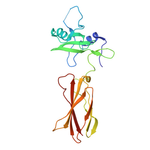
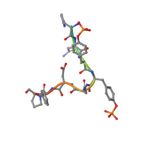
    4JMG, 4JMH

  • PubMed Abstract: 

    Cell signaling depends on dynamic protein-protein interaction (PPI) networks, often assembled through modular domains each interacting with multiple peptide motifs. This complexity raises a conceptual challenge, namely to define whether a particular cellular response requires assembly of the complete PPI network of interest or can be driven by a specific interaction. To address this issue, we designed variants of the Grb2 SH2 domain ("pY-clamps") whose specificity is highly biased toward a single phosphotyrosine (pY) motif among many potential pYXNX Grb2-binding sites. Surprisingly, directing Grb2 predominantly to a single pY site of the Ptpn11/Shp2 phosphatase, but not other sites tested, was sufficient for differentiation of the essential primitive endoderm lineage from embryonic stem cells. Our data suggest that discrete connections within complex PPI networks can underpin regulation of particular biological events. We propose that this directed wiring approach will be of general utility in functionally annotating specific PPIs.


  • Organizational Affiliation

    Department of Biochemistry and Molecular Biology, The University of Chicago, Chicago, IL 60637, USA.


Macromolecules
Find similar proteins by:  (by identity cutoff)  |  3D Structure
Entity ID: 1
MoleculeChains Sequence LengthOrganismDetailsImage
Clamp Shc1_pY239/240202synthetic constructMutation(s): 0 
Entity Groups  
Sequence Clusters30% Identity50% Identity70% Identity90% Identity95% Identity100% Identity
Sequence Annotations
Expand
  • Reference Sequence

Find similar proteins by:  Sequence   |   3D Structure  

Entity ID: 2
MoleculeChains Sequence LengthOrganismDetailsImage
SHC-transforming protein 113Homo sapiensMutation(s): 0 
Gene Names: SHCSHC1SHCA
UniProt & NIH Common Fund Data Resources
Find proteins for P29353 (Homo sapiens)
Explore P29353 
Go to UniProtKB:  P29353
PHAROS:  P29353
GTEx:  ENSG00000160691 
Entity Groups  
Sequence Clusters30% Identity50% Identity70% Identity90% Identity95% Identity100% Identity
UniProt GroupP29353
Sequence Annotations
Expand
  • Reference Sequence
Small Molecules
Modified Residues  1 Unique
IDChains TypeFormula2D DiagramParent
PTR
Query on PTR
B
L-PEPTIDE LINKINGC9 H12 N O6 PTYR
Experimental Data & Validation

Experimental Data

  • Method: X-RAY DIFFRACTION
  • Resolution: 2.41 Å
  • R-Value Free: 0.276 
  • R-Value Work: 0.213 
  • R-Value Observed: 0.216 
  • Space Group: C 2 2 21
Unit Cell:
Length ( Å )Angle ( ˚ )
a = 89.853α = 90
b = 92.19β = 90
c = 59.596γ = 90
Software Package:
Software NamePurpose
PHASESphasing
PHENIXrefinement
HKL-2000data reduction
HKL-2000data scaling

Structure Validation

View Full Validation Report


Entry History 

Deposition Data

Revision History  (Full details and data files)

  • Version 1.0: 2014-04-23
    Type: Initial release
  • Version 1.1: 2014-07-30
    Changes: Database references
  • Version 1.2: 2014-08-06
    Changes: Data collection
  • Version 1.3: 2023-09-20
    Changes: Data collection, Database references, Derived calculations, Refinement description
  • Version 1.4: 2023-12-06
    Changes: Data collection