A Plasmodium falciparum PHIST protein binds the virulence factor PfEMP1 and comigrates to knobs on the host cell surface.
Oberli, A., Slater, L.M., Cutts, E., Brand, F., Mundwiler-Pachlatko, E., Rusch, S., Masik, M.F., Erat, M.C., Beck, H.P., Vakonakis, I.(2014) FASEB J 28: 4420-4433
- PubMed: 24983468
- DOI: https://doi.org/10.1096/fj.14-256057
- Primary Citation of Related Structures:
4JLE - PubMed Abstract:
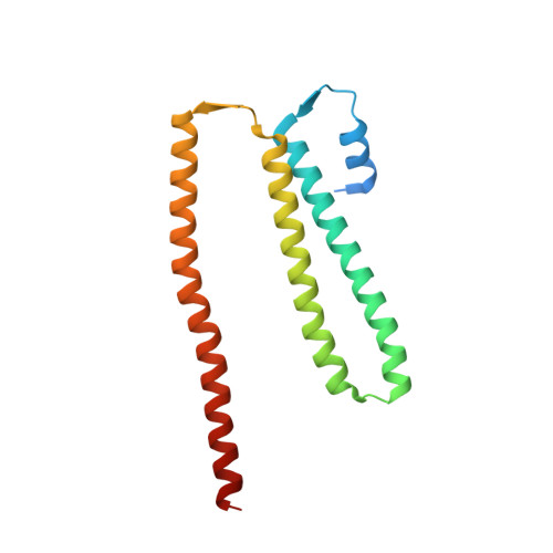
Uniquely among malaria parasites, Plasmodium falciparum-infected erythrocytes (iRBCs) develop membrane protrusions, known as knobs, where the parasite adhesion receptor P. falciparum erythrocyte membrane protein 1 (PfEMP1) clusters. Knob formation and the associated iRBC adherence to host endothelium are directly linked to the severity of malaria and are functional manifestations of protein export from the parasite to the iRBC. A family of exported proteins featuring Plasmodium helical interspersed subtelomeric (PHIST) domains has attracted attention, with members being implicated in host-parasite protein interactions and differentially regulated in severe disease and among parasite isolates. Here, we show that PHIST member PFE1605w binds the PfEMP1 intracellular segment directly with Kd = 5 ± 0.6 μM, comigrates with PfEMP1 during export, and locates in knobs. PHIST variants that do not locate in knobs (MAL8P1.4) or bind PfEMP1 30 times more weakly (PFI1780w) used as controls did not display the same pattern. We resolved the first crystallographic structure of a PHIST protein and derived a partial model of the PHIST-PfEMP1 interaction from nuclear magnetic resonance. We propose that PFE1605w reinforces the PfEMP1-cytoskeletal connection in knobs and discuss the possible role of PHIST proteins as interaction hubs in the parasite exportome.
Organizational Affiliation:
Swiss Tropical and Public Health Institute, Basel, Switzerland; University of Basel, Basel, Switzerland; and.