4JKW

Structure of the extracellular domain of butyrophilin BTN3A1 in complex with Isopentenyl pyrophosphate (IPP)


Experimental Data Snapshot

  • Method: X-RAY DIFFRACTION
  • Resolution: 2.01 Å
  • R-Value Free: 0.268 
  • R-Value Work: 0.229 
  • R-Value Observed: 0.231 

wwPDB Validation   3D Report Full Report


Ligand Structure Quality Assessment 


This is version 1.3 of the entry. See complete history


Literature

Butyrophilin 3A1 binds phosphorylated antigens and stimulates human gamma delta T cells.

Vavassori, S.Kumar, A.Wan, G.S.Ramanjaneyulu, G.S.Cavallari, M.El Daker, S.Beddoe, T.Theodossis, A.Williams, N.K.Gostick, E.Price, D.A.Soudamini, D.U.Voon, K.K.Olivo, M.Rossjohn, J.Mori, L.De Libero, G.

(2013) Nat Immunol 14: 908-916

  • DOI: https://doi.org/10.1038/ni.2665
  • Primary Citation of Related Structures:  
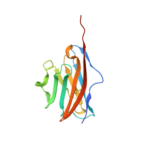
    4JKW, 4K55

  • PubMed Abstract: 

    Human T cells that express a T cell antigen receptor (TCR) containing γ-chain variable region 9 and δ-chain variable region 2 (Vγ9Vδ2) recognize phosphorylated prenyl metabolites as antigens in the presence of antigen-presenting cells but independently of major histocompatibility complex (MHC), the MHC class I-related molecule MR1 and antigen-presenting CD1 molecules. Here we used genetic approaches to identify the molecule that binds and presents phosphorylated antigens. We found that the butyrophilin BTN3A1 bound phosphorylated antigens with low affinity, at a stoichiometry of 1:1, and stimulated mouse T cells with transgenic expression of a human Vγ9Vδ2 TCR. The structures of the BTN3A1 distal domain in complex with host- or microbe-derived phosphorylated antigens had an immunoglobulin-like fold in which the antigens bound in a shallow pocket. Soluble Vγ9Vδ2 TCR interacted specifically with BTN3A1-antigen complexes. Accordingly, BTN3A1 represents an antigen-presenting molecule required for the activation of Vγ9Vδ2 T cells.


  • Organizational Affiliation

    Experimental Immunology, Department of Biomedicine, University Hospital Basel, Basel, Switzerland.


Macromolecules
Find similar proteins by:  (by identity cutoff)  |  3D Structure
Entity ID: 1
MoleculeChains Sequence LengthOrganismDetailsImage
Butyrophilin subfamily 3 member A1124Homo sapiensMutation(s): 0 
Gene Names: BT3.2BTF3BTF4BTF5BTN3A1BTN3A2Butyrophilin subfamily 3 member A1
UniProt & NIH Common Fund Data Resources
Find proteins for O00481 (Homo sapiens)
Explore O00481 
Go to UniProtKB:  O00481
PHAROS:  O00481
GTEx:  ENSG00000026950 
Entity Groups  
Sequence Clusters30% Identity50% Identity70% Identity90% Identity95% Identity100% Identity
UniProt GroupO00481
Sequence Annotations
Expand
  • Reference Sequence
Small Molecules
Ligands 1 Unique
IDChains Name / Formula / InChI Key2D Diagram3D Interactions
IPE
Query on IPE

Download Ideal Coordinates CCD File 
B [auth A]3-METHYLBUT-3-ENYL TRIHYDROGEN DIPHOSPHATE
C5 H12 O7 P2
NUHSROFQTUXZQQ-UHFFFAOYSA-N
Binding Affinity Annotations 
IDSourceBinding Affinity
IPE PDBBind:  4JKW Kd: 6.99e+4 (nM) from 1 assay(s)
Binding MOAD:  4JKW Kd: 6.99e+4 (nM) from 1 assay(s)
Experimental Data & Validation

Experimental Data

  • Method: X-RAY DIFFRACTION
  • Resolution: 2.01 Å
  • R-Value Free: 0.268 
  • R-Value Work: 0.229 
  • R-Value Observed: 0.231 
  • Space Group: P 21 21 21
Unit Cell:
Length ( Å )Angle ( ˚ )
a = 32.717α = 90
b = 36.194β = 90
c = 108.013γ = 90
Software Package:
Software NamePurpose
HKL-2000data collection
PHASERphasing
REFMACrefinement
HKL-2000data reduction
HKL-2000data scaling

Structure Validation

View Full Validation Report


Ligand Structure Quality Assessment 


Entry History 

Deposition Data

Revision History  (Full details and data files)

  • Version 1.0: 2013-07-24
    Type: Initial release
  • Version 1.1: 2013-07-31
    Changes: Structure summary
  • Version 1.2: 2013-08-28
    Changes: Database references
  • Version 1.3: 2013-09-25
    Changes: Database references