4JKN

Mercury Metallated Pseudomonas aeruginosa Azurin at 1.54 A


Experimental Data Snapshot

  • Method: X-RAY DIFFRACTION
  • Resolution: 1.54 Å
  • R-Value Free: 0.239 
  • R-Value Work: 0.211 
  • R-Value Observed: 0.213 

wwPDB Validation   3D Report Full Report


This is version 1.3 of the entry. See complete history


Literature

Mercury metallation of the copper protein azurin and structural insight into possible heavy metal reactivity.

Zampino, A.P.Masters, F.M.Bladholm, E.L.Panzner, M.J.Berry, S.M.Leeper, T.C.Ziegler, C.J.

(2014) J Inorg Biochem 141: 152-160

  • DOI: https://doi.org/10.1016/j.jinorgbio.2014.09.003
  • Primary Citation of Related Structures:  
    4JKN

  • PubMed Abstract: 

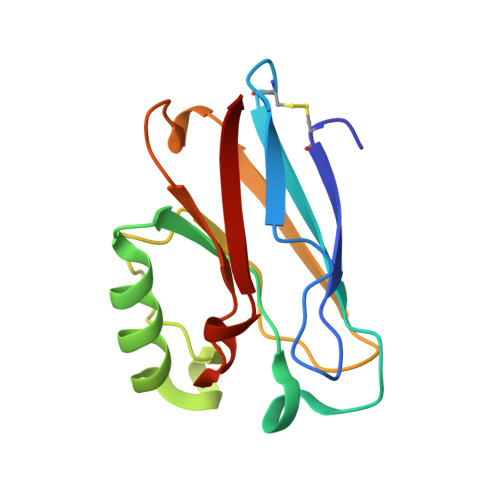
    Mercury(II) metallation of Pseudomonas aeruginosa azurin has been characterized structurally and biochemically. The X-ray crystal structure at 1.5Å of mercury(II) metallated azurin confirms the coordination of mercury at the copper binding active site and a second surface site. These findings are further validated by NMR, Matrix-assisted laser desorption/ionization spectrometry (MALDI), and UV-visible spectroscopic methods indicating copper displacement from the wild-type protein. Bioinformatic analysis has identified homologous human protein domains computationally, and compared them to the structure of azurin, providing a model for human mercury interactions. Study of the mercury-azurin adduct, in combination with other known examples of protein-heavy metal interactions, could provide further insight into the chemical mechanisms of toxicological interactions, leading toward a global understanding of the biological speciation of toxic heavy metals.


  • Organizational Affiliation

    Department of Chemistry, The University of Akron, Akron, OH 44325, USA.


Macromolecules
Find similar proteins by:  (by identity cutoff)  |  3D Structure
Entity ID: 1
MoleculeChains Sequence LengthOrganismDetailsImage
Azurin
A, B, C, D
128Pseudomonas aeruginosa PAO1Mutation(s): 0 
Gene Names: azuazu PA4922PA4922
EC: 1.20.9.1
UniProt
Find proteins for P00282 (Pseudomonas aeruginosa (strain ATCC 15692 / DSM 22644 / CIP 104116 / JCM 14847 / LMG 12228 / 1C / PRS 101 / PAO1))
Explore P00282 
Go to UniProtKB:  P00282
Entity Groups  
Sequence Clusters30% Identity50% Identity70% Identity90% Identity95% Identity100% Identity
UniProt GroupP00282
Sequence Annotations
Expand
  • Reference Sequence
Small Molecules
Ligands 3 Unique
IDChains Name / Formula / InChI Key2D Diagram3D Interactions
HG
Query on HG

Download Ideal Coordinates CCD File 
E [auth A],
F [auth A],
I [auth B],
K [auth C],
L [auth D]
MERCURY (II) ION
Hg
BQPIGGFYSBELGY-UHFFFAOYSA-N
SO4
Query on SO4

Download Ideal Coordinates CCD File 
Q [auth D]SULFATE ION
O4 S
QAOWNCQODCNURD-UHFFFAOYSA-L
NO3
Query on NO3

Download Ideal Coordinates CCD File 
G [auth A]
H [auth A]
J [auth B]
M [auth D]
N [auth D]
G [auth A],
H [auth A],
J [auth B],
M [auth D],
N [auth D],
O [auth D],
P [auth D]
NITRATE ION
N O3
NHNBFGGVMKEFGY-UHFFFAOYSA-N
Experimental Data & Validation

Experimental Data

  • Method: X-RAY DIFFRACTION
  • Resolution: 1.54 Å
  • R-Value Free: 0.239 
  • R-Value Work: 0.211 
  • R-Value Observed: 0.213 
  • Space Group: P 21 21 21
Unit Cell:
Length ( Å )Angle ( ˚ )
a = 56.483α = 90
b = 79.228β = 90
c = 109.576γ = 90
Software Package:
Software NamePurpose
APEXdata collection
PHASESphasing
PHENIXrefinement
SAINTdata reduction
APEXdata reduction

Structure Validation

View Full Validation Report


Entry History 

Deposition Data

Revision History  (Full details and data files)

  • Version 1.0: 2014-02-19
    Type: Initial release
  • Version 1.1: 2015-02-04
    Changes: Database references
  • Version 1.2: 2018-04-04
    Changes: Data collection
  • Version 1.3: 2023-09-20
    Changes: Data collection, Database references, Derived calculations, Refinement description