Development of novel molecular probes of the Rio1 atypical protein kinase.
Mielecki, M., Krawiec, K., Kiburu, I., Grzelak, K., Zagorski, W., Kierdaszuk, B., Kowa, K., Fokt, I., Szymanski, S., Swierk, P., Szeja, W., Priebe, W., Lesyng, B., Laronde-Leblanc, N.(2013) Biochim Biophys Acta 1834: 1292-1301
- PubMed: 23523885
- DOI: https://doi.org/10.1016/j.bbapap.2013.03.012
- Primary Citation of Related Structures:
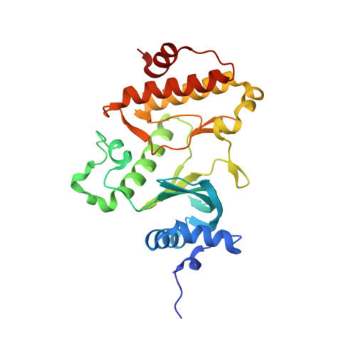
4JIN - PubMed Abstract:
The RIO kinases are essential protein factors required for the synthesis of new ribosomes in eukaryotes. Conserved in archaeal organisms as well, RIO kinases are among the most ancient of protein kinases. Their exact molecular mechanisms are under investigation and progress of this research would be significantly improved with the availability of suitable molecular probes that selectively block RIO kinases. RIO kinases contain a canonical eukaryotic protein kinase fold, but also display several unusual structural features that potentially create opportunity for the design of selective inhibitors. In an attempt to identify structural leads to target the RIO kinases, a series of pyridine caffeic acid benzyl amides (CABA) were tested for their ability to inhibit the autophosphorylation activity of Archeaoglobus fulgidus Rio1 (AfRio1). Screening of a small library of CABA molecules resulted in the identification of four compounds that measurably inhibited AfRio1 activity. Additional biochemical characterization of binding and inhibition activity of these compounds demonstrated an ATP competitive inhibition mode, and allowed identification of the functional groups that result in the highest binding affinity. In addition, docking of the compound to the structure of Rio1 and determination of the X-ray crystal structure of a model compound (WP1086) containing the desired functional groups allowed detailed analysis of the interactions between these compounds and the enzyme. Furthermore, the X-ray crystal structure demonstrated that these compounds stabilize an inactive form of the enzyme. Taken together, these results provide an important step in identification of a scaffold for the design of selective molecular probes to study molecular mechanisms of Rio1 kinases in vitro and in vivo. In addition, it provides a rationale for the future design of potent inhibitors with drug-like properties targeting an inactive form of the enzyme. This article is part of a Special Issue entitled: Inhibitors of Protein Kinases (2012).
Organizational Affiliation:
Polish Academy of Sciences, Institute of Biochemistry and Biophysics, Protein Biosynthesis Department, Warsaw, Poland.