


X-ray crystal structure of 3-oxoacyl-[acyl-carrier-protein] synthase 2 from Rickettsia rickettsii
Fairman, J.W., Abendroth, J.A., Edwards, T.E., Lorimer, D.To be published.
Experimental Data Snapshot
wwPDB Validation 3D Report Full Report

Entity ID: 1 | |||||
|---|---|---|---|---|---|
| Molecule | Chains | Sequence Length | Organism | Details | Image |
| 3-oxoacyl-[acyl-carrier-protein] synthase 2 | 437 | Rickettsia rickettsii str. 'Sheila Smith | Mutation(s): 0 Gene Names: A1G_06510 EC: 2.3.1.179 | 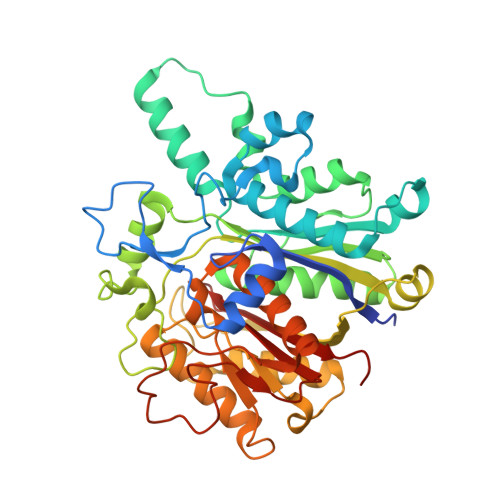 | |
UniProt | |||||
Find proteins for A0A0H3AY56 (Rickettsia rickettsii (strain Sheila Smith)) Explore A0A0H3AY56 Go to UniProtKB: A0A0H3AY56 | |||||
Entity Groups | |||||
| Sequence Clusters | 30% Identity50% Identity70% Identity90% Identity95% Identity100% Identity | ||||
| UniProt Group | A0A0H3AY56 | ||||
Sequence AnnotationsExpand | |||||
| |||||
| Ligands 3 Unique | |||||
|---|---|---|---|---|---|
| ID | Chains | Name / Formula / InChI Key | 2D Diagram | 3D Interactions | |
| EDO Query on EDO | D [auth A] | 1,2-ETHANEDIOL C2 H6 O2 LYCAIKOWRPUZTN-UHFFFAOYSA-N |  | ||
| K Query on K | C [auth A], F [auth B] | POTASSIUM ION K NPYPAHLBTDXSSS-UHFFFAOYSA-N |  | ||
| NA Query on NA | E [auth A] | SODIUM ION Na FKNQFGJONOIPTF-UHFFFAOYSA-N |  | ||
| Length ( Å ) | Angle ( ˚ ) |
|---|---|
| a = 73.17 | α = 90 |
| b = 62.74 | β = 112.08 |
| c = 95.43 | γ = 90 |
| Software Name | Purpose |
|---|---|
| XSCALE | data scaling |
| PHASER | phasing |
| REFMAC | refinement |
| PDB_EXTRACT | data extraction |
RCSB PDB (citation) is hosted by
RCSB PDB is a member of the

