4JBL

Crystal structure of O-Acetyl Serine Sulfhydrylase from Entamoeba histolytica in complex with Methionine


Experimental Data Snapshot

  • Method: X-RAY DIFFRACTION
  • Resolution: 2.00 Å
  • R-Value Free: 0.225 
  • R-Value Work: 0.178 
  • R-Value Observed: 0.181 

wwPDB Validation   3D Report Full Report


This is version 1.3 of the entry. See complete history


Literature

Molecular basis of ligand recognition by OASS from E. histolytica: insights from structural and molecular dynamics simulation studies

Raj, I.Mazumder, M.Gourinath, S.

(2013) Biochim Biophys Acta 1830: 4573-4583

  • DOI: https://doi.org/10.1016/j.bbagen.2013.05.041
  • Primary Citation of Related Structures:  
    4IL5, 4JBL, 4JBN

  • PubMed Abstract: 

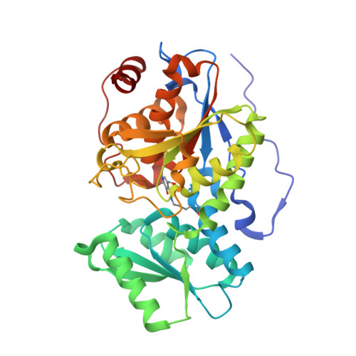
    O-acetyl serine sulfhydrylase (OASS) is a pyridoxal phosphate (PLP) dependent enzyme catalyzing the last step of the cysteine biosynthetic pathway. Here we analyze and investigate the factors responsible for recognition and different conformational changes accompanying the binding of various ligands to OASS.


  • Organizational Affiliation

    School of Life Sciences, Jawaharlal Nehru University, New Delhi, India.


Macromolecules
Find similar proteins by:  (by identity cutoff)  |  3D Structure
Entity ID: 1
MoleculeChains Sequence LengthOrganismDetailsImage
Cysteine synthase
A, B
339Entamoeba histolyticaMutation(s): 0 
Gene Names: EhOASS
EC: 2.5.1.47
UniProt
Find proteins for O15570 (Entamoeba histolytica)
Explore O15570 
Go to UniProtKB:  O15570
Entity Groups  
Sequence Clusters30% Identity50% Identity70% Identity90% Identity95% Identity100% Identity
UniProt GroupO15570
Sequence Annotations
Expand
  • Reference Sequence
Small Molecules
Modified Residues  1 Unique
IDChains TypeFormula2D DiagramParent
LLP
Query on LLP
A, B
L-PEPTIDE LINKINGC14 H22 N3 O7 PLYS
Binding Affinity Annotations 
IDSourceBinding Affinity
MET PDBBind:  4JBL Kd: 5.40e+5 (nM) from 1 assay(s)
Binding MOAD:  4JBL Kd: 5.40e+5 (nM) from 1 assay(s)
Experimental Data & Validation

Experimental Data

  • Method: X-RAY DIFFRACTION
  • Resolution: 2.00 Å
  • R-Value Free: 0.225 
  • R-Value Work: 0.178 
  • R-Value Observed: 0.181 
  • Space Group: P 41
Unit Cell:
Length ( Å )Angle ( ˚ )
a = 80.417α = 90
b = 80.417β = 90
c = 112.235γ = 90
Software Package:
Software NamePurpose
SCALEPACKdata scaling
REFMACrefinement
PDB_EXTRACTdata extraction
MAR345dtbdata collection
AUTOMARdata reduction
MOLREPphasing

Structure Validation

View Full Validation Report


Entry History 

Deposition Data

Revision History  (Full details and data files)

  • Version 1.0: 2013-12-04
    Type: Initial release
  • Version 1.1: 2023-11-08
    Changes: Data collection, Database references, Derived calculations, Refinement description
  • Version 1.2: 2023-12-06
    Changes: Data collection
  • Version 1.3: 2024-03-20
    Changes: Source and taxonomy, Structure summary