4J7I

SET7/9Y335F in complex with TAF10 peptide and AdoHcy


Experimental Data Snapshot

  • Method: X-RAY DIFFRACTION
  • Resolution: 2.56 Å
  • R-Value Free: 0.257 
  • R-Value Work: 0.202 
  • R-Value Observed: 0.205 

wwPDB Validation   3D Report Full Report


Ligand Structure Quality Assessment 


This is version 1.1 of the entry. See complete history


Literature

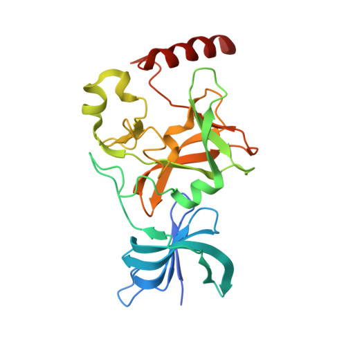
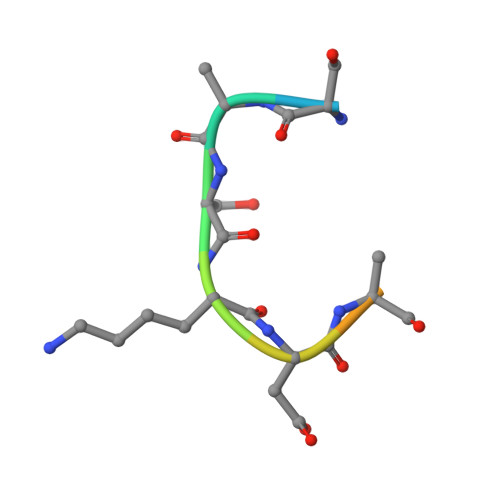
Conservation and functional importance of carbon-oxygen hydrogen bonding in AdoMet-dependent methyltransferases.

Horowitz, S.Dirk, L.M.Yesselman, J.D.Nimtz, J.S.Adhikari, U.Mehl, R.A.Scheiner, S.Houtz, R.L.Al-Hashimi, H.M.Trievel, R.C.

(2013) J Am Chem Soc 135: 15536-15548

  • DOI: https://doi.org/10.1021/ja407140k
  • Primary Citation of Related Structures:  
    4J7I, 4J83, 4J8O

  • PubMed Abstract: 

    S-adenosylmethionine (AdoMet)-based methylation is integral to metabolism and signaling. AdoMet-dependent methyltransferases belong to multiple distinct classes and share a catalytic mechanism that arose through convergent evolution; however, fundamental determinants underlying this shared methyl transfer mechanism remain undefined. A survey of high-resolution crystal structures reveals that unconventional carbon-oxygen (CH···O) hydrogen bonds coordinate the AdoMet methyl group in different methyltransferases irrespective of their class, active site structure, or cofactor binding conformation. Corroborating these observations, quantum chemistry calculations demonstrate that these charged interactions formed by the AdoMet sulfonium cation are stronger than typical CH···O hydrogen bonds. Biochemical and structural studies using a model lysine methyltransferase and an active site mutant that abolishes CH···O hydrogen bonding to AdoMet illustrate that these interactions are important for high-affinity AdoMet binding and transition-state stabilization. Further, crystallographic and NMR dynamics experiments of the wild-type enzyme demonstrate that the CH···O hydrogen bonds constrain the motion of the AdoMet methyl group, potentially facilitating its alignment during catalysis. Collectively, the experimental findings with the model methyltransferase and structural survey imply that methyl CH···O hydrogen bonding represents a convergent evolutionary feature of AdoMet-dependent methyltransferases, mediating a universal mechanism for methyl transfer.


  • Organizational Affiliation

    Howard Hughes Medical Institute , Ann Arbor, Michigan 48109, United States.


Macromolecules
Find similar proteins by:  (by identity cutoff)  |  3D Structure
Entity ID: 1
MoleculeChains Sequence LengthOrganismDetailsImage
Histone-lysine N-methyltransferase SETD7261Homo sapiensMutation(s): 1 
Gene Names: SETD7KIAA1717KMT7SET7SET9
EC: 2.1.1.43
UniProt & NIH Common Fund Data Resources
Find proteins for Q8WTS6 (Homo sapiens)
Explore Q8WTS6 
Go to UniProtKB:  Q8WTS6
PHAROS:  Q8WTS6
GTEx:  ENSG00000145391 
Entity Groups  
Sequence Clusters30% Identity50% Identity70% Identity90% Identity95% Identity100% Identity
UniProt GroupQ8WTS6
Sequence Annotations
Expand
  • Reference Sequence

Find similar proteins by:  Sequence   |   3D Structure  

Entity ID: 2
MoleculeChains Sequence LengthOrganismDetailsImage
Transcription initiation factor TFIID subunit 1011Homo sapiensMutation(s): 0 
UniProt & NIH Common Fund Data Resources
Find proteins for Q12962 (Homo sapiens)
Explore Q12962 
Go to UniProtKB:  Q12962
PHAROS:  Q12962
GTEx:  ENSG00000166337 
Entity Groups  
Sequence Clusters30% Identity50% Identity70% Identity90% Identity95% Identity100% Identity
UniProt GroupQ12962
Sequence Annotations
Expand
  • Reference Sequence
Small Molecules
Ligands 1 Unique
IDChains Name / Formula / InChI Key2D Diagram3D Interactions
SAH
Query on SAH

Download Ideal Coordinates CCD File 
C [auth A]S-ADENOSYL-L-HOMOCYSTEINE
C14 H20 N6 O5 S
ZJUKTBDSGOFHSH-WFMPWKQPSA-N
Binding Affinity Annotations 
IDSourceBinding Affinity
SAH BindingDB:  4J7I IC50: min: 3.00e+4, max: 4.70e+4 (nM) from 2 assay(s)
Experimental Data & Validation

Experimental Data

  • Method: X-RAY DIFFRACTION
  • Resolution: 2.56 Å
  • R-Value Free: 0.257 
  • R-Value Work: 0.202 
  • R-Value Observed: 0.205 
  • Space Group: P 32 2 1
Unit Cell:
Length ( Å )Angle ( ˚ )
a = 82.305α = 90
b = 82.305β = 90
c = 95.268γ = 120
Software Package:
Software NamePurpose
HKL-2000data collection
MOLREPphasing
REFMACrefinement
HKL-2000data reduction
HKL-2000data scaling

Structure Validation

View Full Validation Report


Ligand Structure Quality Assessment 


Entry History 

Deposition Data

Revision History  (Full details and data files)

  • Version 1.0: 2014-01-08
    Type: Initial release
  • Version 1.1: 2024-02-28
    Changes: Data collection, Database references, Derived calculations