4J4M

Crystal structure of TM-1, a Trimeresurus mucrosquamatus venom metalloproteinase


Experimental Data Snapshot

  • Method: X-RAY DIFFRACTION
  • Resolution: 1.80 Å
  • R-Value Free: 0.207 
  • R-Value Work: 0.184 
  • R-Value Observed: 0.188 

wwPDB Validation   3D Report Full Report


This is version 1.1 of the entry. See complete history


Literature

Crystal structure of a Trimeresurus mucrosquamatus venom metalloproteinase providing new insights into the inhibition by endogenous tripeptide inhibitors.

Chou, T.L.Wu, C.H.Huang, K.F.Wang, A.H.

(2013) Toxicon 71C: 140-146

  • DOI: https://doi.org/10.1016/j.toxicon.2013.05.009
  • Primary Citation of Related Structures:  
    4J4M

  • PubMed Abstract: 

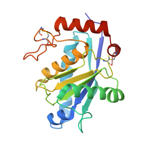
    The crystal structure of TM-1, a P-I class snake-venom metalloproteinase (SVMP) from the Trimeresurus mucrosquamatus venom, was determined at 1.8-Å resolution. The structure exhibits the typical feature of SVMPs and is stabilized by three disulfide linkages. The active site shows a deep S1' substrate-binding pocket limited by the non-conserved Pro174 at the bottom. Further comparisons with other SVMPs suggest that the deep S1' site of TM-1 correlates with its high inhibition sensitivity to the endogenous tripeptide inhibitors. Proteolytic specificity analysis revealed that TM-1 prefers substrates having a moderate-size and hydrophobic residue at the P1' position, consistent with our structural observation.


  • Organizational Affiliation

    Institute of Biochemical Sciences, National Taiwan University, Taipei 10617, Taiwan.


Macromolecules
Find similar proteins by:  (by identity cutoff)  |  3D Structure
Entity ID: 1
MoleculeChains Sequence LengthOrganismDetailsImage
zinc-dependent metalloproteinase
A, B
202Protobothrops mucrosquamatusMutation(s): 0 
EC: 3.4.24.53
UniProt
Find proteins for U3KRG1 (Protobothrops mucrosquamatus)
Explore U3KRG1 
Go to UniProtKB:  U3KRG1
Entity Groups  
Sequence Clusters30% Identity50% Identity70% Identity90% Identity95% Identity100% Identity
UniProt GroupU3KRG1
Sequence Annotations
Expand
  • Reference Sequence
Experimental Data & Validation

Experimental Data

  • Method: X-RAY DIFFRACTION
  • Resolution: 1.80 Å
  • R-Value Free: 0.207 
  • R-Value Work: 0.184 
  • R-Value Observed: 0.188 
  • Space Group: P 61
Unit Cell:
Length ( Å )Angle ( ˚ )
a = 80.766α = 90
b = 80.766β = 90
c = 120.451γ = 120
Software Package:
Software NamePurpose
HKL-2000data collection
CNSrefinement
HKL-2000data reduction
HKL-2000data scaling
CNSphasing

Structure Validation

View Full Validation Report


Entry History 

Deposition Data

Revision History  (Full details and data files)

  • Version 1.0: 2013-07-10
    Type: Initial release
  • Version 1.1: 2023-11-08
    Changes: Data collection, Database references, Derived calculations, Refinement description