4J3U

Crystal structure of barley limit dextrinase in complex with maltosyl-S-betacyclodextrin


Experimental Data Snapshot

  • Method: X-RAY DIFFRACTION
  • Resolution: 1.70 Å
  • R-Value Free: 0.218 
  • R-Value Work: 0.172 
  • R-Value Observed: 0.175 

wwPDB Validation   3D Report Full Report


This is version 2.1 of the entry. See complete history


Literature

Oligosaccharide and substrate binding in the starch debranching enzyme barley limit dextrinase

Moeller, M.S.Windahl, M.S.Sim, L.Bjstrup, M.Abou Hachem, M.Hindsgaul, O.Palcic, M.Svensson, B.Henriksen, A.

(2015) J Mol Biol 427: 1263-1277

  • DOI: https://doi.org/10.1016/j.jmb.2014.12.019
  • Primary Citation of Related Structures:  
    4J3S, 4J3T, 4J3U, 4J3V, 4J3W, 4J3X

  • PubMed Abstract: 

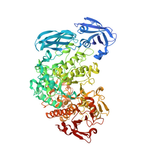
    Complete hydrolytic degradation of starch requires hydrolysis of both the α-1,4- and α-1,6-glucosidic bonds in amylopectin. Limit dextrinase (LD) is the only endogenous barley enzyme capable of hydrolyzing the α-1,6-glucosidic bond during seed germination, and impaired LD activity inevitably reduces the maltose and glucose yields from starch degradation. Crystal structures of barley LD and active-site mutants with natural substrates, products and substrate analogues were sought to better understand the facets of LD-substrate interactions that confine high activity of LD to branched maltooligosaccharides. For the first time, an intact α-1,6-glucosidically linked substrate spanning the active site of a LD or pullulanase has been trapped and characterized by crystallography. The crystal structure reveals both the branch and main-chain binding sites and is used to suggest a mechanism for nucleophilicity enhancement in the active site. The substrate, product and analogue complexes were further used to outline substrate binding subsites and substrate binding restraints and to suggest a mechanism for avoidance of dual α-1,6- and α-1,4-hydrolytic activity likely to be a biological necessity during starch synthesis.


  • Organizational Affiliation

    Carlsberg Laboratory, Gamle Carlsberg Vej 10, DK-1799 Copenhagen V, Denmark; Enzyme and Protein Chemistry, Department of Systems Biology, Technical University of Denmark, Søltofts Plads, Building 224, DK-2800 Kongens Lyngby, Denmark.


Macromolecules
Find similar proteins by:  (by identity cutoff)  |  3D Structure
Entity ID: 1
MoleculeChains Sequence LengthOrganismDetailsImage
Limit dextrinase
A, B
905Hordeum vulgareMutation(s): 0 
EC: 3.2.1.41
UniProt
Find proteins for Q9FYY0 (Hordeum vulgare)
Explore Q9FYY0 
Go to UniProtKB:  Q9FYY0
Entity Groups  
Sequence Clusters30% Identity50% Identity70% Identity90% Identity95% Identity100% Identity
UniProt GroupQ9FYY0
Sequence Annotations
Expand
  • Reference Sequence
Oligosaccharides

Help

Entity ID: 2
MoleculeChains Length2D Diagram Glycosylation3D Interactions
Cyclic alpha-D-glucopyranose-(1-4)-alpha-D-glucopyranose-(1-4)-alpha-D-glucopyranose-(1-4)-alpha-D-glucopyranose-(1-4)-alpha-D-glucopyranose-(1-4)-alpha-D-glucopyranose-(1-4)-[alpha-D-glucopyranose-(1-4)-alpha-D-glucopyranose-(1-1)]6-thio-alpha-D-glucopyranose
C, D
9N/AN/A
Glycosylation Resources
GlyTouCan:  G65588TB
GlyCosmos:  G65588TB
Small Molecules
Ligands 3 Unique
IDChains Name / Formula / InChI Key2D Diagram3D Interactions
IOD
Query on IOD

Download Ideal Coordinates CCD File 
BA [auth B]
CA [auth B]
DA [auth B]
E [auth A]
EA [auth B]
BA [auth B],
CA [auth B],
DA [auth B],
E [auth A],
EA [auth B],
F [auth A],
FA [auth B],
G [auth A],
GA [auth B],
H [auth A],
HA [auth B],
I [auth A],
IA [auth B],
J [auth A],
JA [auth B],
K [auth A],
KA [auth B],
L [auth A],
LA [auth B],
M [auth A],
MA [auth B],
N [auth A],
O [auth A],
P [auth A],
Q [auth A],
R [auth A],
S [auth A],
T [auth A],
U [auth A],
V [auth A],
W [auth A],
X [auth A]
IODIDE ION
I
XMBWDFGMSWQBCA-UHFFFAOYSA-M
CA
Query on CA

Download Ideal Coordinates CCD File 
NA [auth B],
OA [auth B],
Y [auth A],
Z [auth A]
CALCIUM ION
Ca
BHPQYMZQTOCNFJ-UHFFFAOYSA-N
CL
Query on CL

Download Ideal Coordinates CCD File 
AA [auth A],
PA [auth B]
CHLORIDE ION
Cl
VEXZGXHMUGYJMC-UHFFFAOYSA-M
Experimental Data & Validation

Experimental Data

  • Method: X-RAY DIFFRACTION
  • Resolution: 1.70 Å
  • R-Value Free: 0.218 
  • R-Value Work: 0.172 
  • R-Value Observed: 0.175 
  • Space Group: C 1 2 1
Unit Cell:
Length ( Å )Angle ( ˚ )
a = 195.43α = 90
b = 84.61β = 119.91
c = 121.79γ = 90
Software Package:
Software NamePurpose
XSCALEdata scaling
REFMACrefinement
PDB_EXTRACTdata extraction
XDSdata reduction
MOLREPphasing

Structure Validation

View Full Validation Report


Entry History 

Deposition Data

Revision History  (Full details and data files)

  • Version 1.0: 2014-02-12
    Type: Initial release
  • Version 1.1: 2015-10-14
    Changes: Database references
  • Version 1.2: 2018-05-30
    Changes: Data collection
  • Version 2.0: 2020-07-29
    Type: Remediation
    Reason: Carbohydrate remediation
    Changes: Atomic model, Data collection, Database references, Derived calculations, Structure summary
  • Version 2.1: 2023-11-08
    Changes: Data collection, Database references, Refinement description, Structure summary