A Molecular Switch Governs the Interaction between the Human Complement Protease C1s and Its Substrate, Complement C4.
Perry, A.J., Wijeyewickrema, L.C., Wilmann, P.G., Gunzburg, M.J., D'Andrea, L., Irving, J.A., Pang, S.S., Duncan, R.C., Wilce, J.A., Whisstock, J.C., Pike, R.N.(2013) J Biol Chem 288: 15821-15829
- PubMed: 23592783
- DOI: https://doi.org/10.1074/jbc.M113.464545
- Primary Citation of Related Structures:
4J1Y - PubMed Abstract:
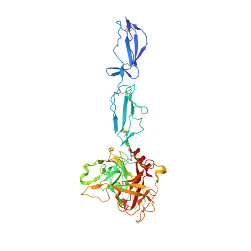
The complement system is an ancient innate immune defense pathway that plays a front line role in eliminating microbial pathogens. Recognition of foreign targets by antibodies drives sequential activation of two serine proteases, C1r and C1s, which reside within the complement Component 1 (C1) complex. Active C1s propagates the immune response through its ability to bind and cleave the effector molecule complement Component 4 (C4). Currently, the precise structural and biochemical basis for the control of the interaction between C1s and C4 is unclear. Here, using surface plasmon resonance, we show that the transition of the C1s zymogen to the active form is essential for C1s binding to C4. To understand this, we determined the crystal structure of a zymogen C1s construct (comprising two complement control protein (CCP) domains and the serine protease (SP) domain). These data reveal that two loops (492-499 and 573-580) in the zymogen serine protease domain adopt a conformation that would be predicted to sterically abrogate C4 binding. The transition from zymogen to active C1s repositions both loops such that they would be able to interact with sulfotyrosine residues on C4. The structure also shows the junction of the CCP1 and CCP2 domains of C1s for the first time, yielding valuable information about the exosite for C4 binding located at this position. Together, these data provide a structural explanation for the control of the interaction with C1s and C4 and, furthermore, point to alternative strategies for developing therapeutic approaches for controlling activation of the complement cascade.
Organizational Affiliation:
Department of Biochemistry and Molecular Biology, Monash University, Clayton, Melbourne, Victoria 3800, Australia.