Structure determination of the functional domain interaction of a chimeric nonribosomal peptide synthetase from a challenging crystal with noncrystallographic translational symmetry.
Sundlov, J.A., Gulick, A.M.(2013) Acta Crystallogr D Biol Crystallogr 69: 1482-1492
- PubMed: 23897471
- DOI: https://doi.org/10.1107/S0907444913009372
- Primary Citation of Related Structures:
4IZ6 - PubMed Abstract:
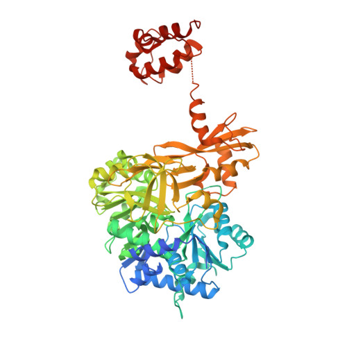
The nonribosomal peptide synthetases (NRPSs) are a family of modular proteins that contain multiple catalytic domains joined in a single protein. Together, these domains work to produce chemically diverse peptides, including compounds with antibiotic activity or that play a role in iron acquisition. Understanding the structural mechanisms that govern the domain interactions has been a long-standing goal. During NRPS synthesis, amino-acid substrates are loaded onto integrated carrier protein domains through the activity of NRPS adenylation domains. The structures of two adenylation domain-carrier protein domain complexes have recently been determined in an effort that required the use of a mechanism-based inhibitor to trap the domain interaction. Here, the continued analysis of these proteins is presented, including a higher resolution structure of an engineered di-domain protein containing the EntE adenylation domain fused with the carrier protein domain of its partner EntB. The protein crystallized in a novel space group in which molecular replacement and refinement were challenged by noncrystallographic pseudo-translational symmetry. The structure determination and how the molecular packing impacted the diffraction intensities are reported. Importantly, the structure illustrates that in this new crystal form the functional interface between the adenylation domain and the carrier protein domain remains the same as that observed previously. At a resolution that allows inclusion of water molecules, additional interactions are observed between the two protein domains and between the protein and its ligands. In particular, a highly solvated region that surrounds the carrier protein cofactor is described.
Organizational Affiliation:
Hauptman-Woodward Medical Research Institute and Department of Structural Biology, University at Buffalo, 700 Ellicott Street, Buffalo, NY 14203, USA.