


Structure of DNA-binding domain of the response regulator SaeR from Staphylococcus epidermidis
Chen, Y.R., Chen, S.C., Yang, C.S., Kuan, S.M., Liu, Y.H., Chen, Y.To be published.
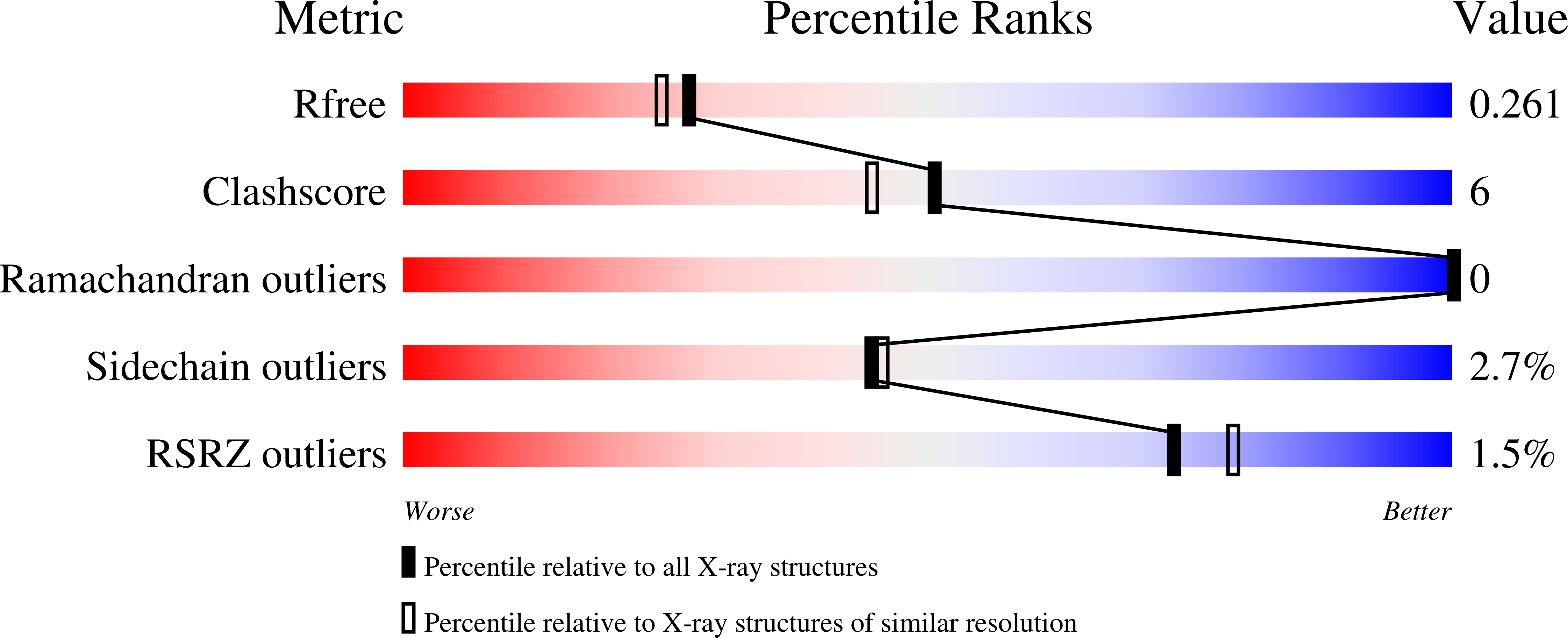
Experimental Data Snapshot
wwPDB Validation 3D Report Full Report
Entity ID: 1 | |||||
|---|---|---|---|---|---|
| Molecule | Chains | Sequence Length | Organism | Details | Image |
| Response regulator SaeR | 110 | Staphylococcus epidermidis ATCC 12228 | Mutation(s): 1 Gene Names: saeR, SE_0479 |  | |
UniProt | |||||
Find proteins for Q8CQ17 (Staphylococcus epidermidis (strain ATCC 12228 / FDA PCI 1200)) Explore Q8CQ17 Go to UniProtKB: Q8CQ17 | |||||
Entity Groups | |||||
| Sequence Clusters | 30% Identity50% Identity70% Identity90% Identity95% Identity100% Identity | ||||
| UniProt Group | Q8CQ17 | ||||
Sequence AnnotationsExpand | |||||
| |||||
| Length ( Å ) | Angle ( ˚ ) |
|---|---|
| a = 34.201 | α = 90 |
| b = 53.784 | β = 90 |
| c = 111.658 | γ = 90 |
| Software Name | Purpose |
|---|---|
| HKL-2000 | data collection |
| PHASER | phasing |
| PHENIX | refinement |
| HKL-2000 | data reduction |
| HKL-2000 | data scaling |
RCSB PDB (citation) is hosted by
RCSB PDB is a member of the

