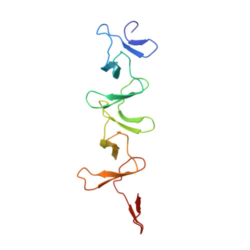
Structural and functional insights into peptidoglycan access for the lytic amidase LytA of Streptococcus pneumoniae.
Mellroth, P., Sandalova, T., Kikhney, A., Vilaplana, F., Hesek, D., Lee, M., Mobashery, S., Normark, S., Svergun, D., Henriques-Normark, B., Achour, A.(2014) mBio 5: e01120-e01113
- PubMed: 24520066
- DOI: https://doi.org/10.1128/mBio.01120-13
- Primary Citation of Related Structures:
4IVV, 4IWT - PubMed Abstract:
The cytosolic N-acetylmuramoyl-l-alanine amidase LytA protein of Streptococcus pneumoniae, which is released by bacterial lysis, associates with the cell wall via its choline-binding motif. During exponential growth, LytA accesses its peptidoglycan substrate to cause lysis only when nascent peptidoglycan synthesis is stalled by nutrient starvation or β-lactam antibiotics. Here we present three-dimensional structures of LytA and establish the requirements for substrate binding and catalytic activity. The solution structure of the full-length LytA dimer reveals a peculiar fold, with the choline-binding domains forming a rigid V-shaped scaffold and the relatively more flexible amidase domains attached in a trans position. The 1.05-Å crystal structure of the amidase domain reveals a prominent Y-shaped binding crevice composed of three contiguous subregions, with a zinc-containing active site localized at the bottom of the branch point. Site-directed mutagenesis was employed to identify catalytic residues and to investigate the relative impact of potential substrate-interacting residues lining the binding crevice for the lytic activity of LytA. In vitro activity assays using defined muropeptide substrates reveal that LytA utilizes a large substrate recognition interface and requires large muropeptide substrates with several connected saccharides that interact with all subregions of the binding crevice for catalysis. We hypothesize that the substrate requirements restrict LytA to the sites on the cell wall where nascent peptidoglycan synthesis occurs.