4IV9

Structure of the Flavoprotein Tryptophan-2-Monooxygenase


Experimental Data Snapshot

  • Method: X-RAY DIFFRACTION
  • Resolution: 1.95 Å
  • R-Value Free: 0.245 
  • R-Value Work: 0.187 
  • R-Value Observed: 0.190 

wwPDB Validation   3D Report Full Report


Ligand Structure Quality Assessment 


This is version 1.2 of the entry. See complete history


Literature

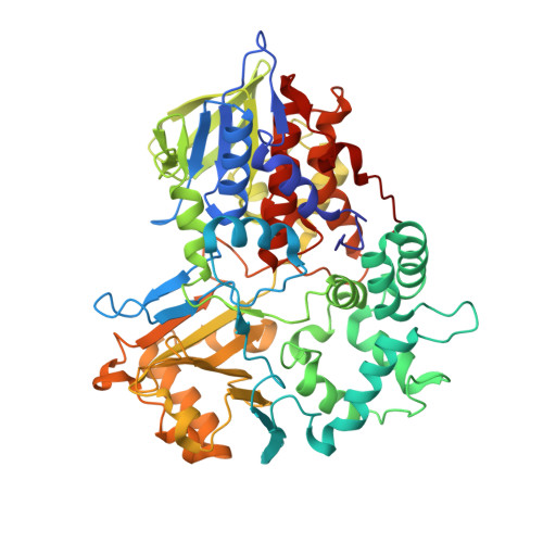
Structure of the flavoprotein tryptophan 2-monooxygenase, a key enzyme in the formation of galls in plants.

Gaweska, H.M.Taylor, A.B.Hart, P.J.Fitzpatrick, P.F.

(2013) Biochemistry 52: 2620-2626

  • DOI: https://doi.org/10.1021/bi4001563
  • Primary Citation of Related Structures:  
    4IV9

  • PubMed Abstract: 

    The flavoprotein tryptophan 2-monooxygenase catalyzes the oxidative decarboxylation of tryptophan to yield indole-3-acetamide. This is the initial step in the biosynthesis of the plant growth hormone indole-acetic acid by bacterial pathogens that cause crown gall and related diseases. The structure of the enzyme from Pseudomonas savastanoi has been determined by X-ray diffraction methods to a resolution of 1.95 Å. The overall structure of the protein shows that it has the same fold as members of the monoamine oxidase family of flavoproteins, with the greatest similarities to the l-amino acid oxidases. The location of bound indole-3-acetamide in the active site allows identification of residues responsible for substrate binding and specificity. Two residues in the enzyme are conserved in all members of the monoamine oxidase family, Lys365 and Trp466. The K365M mutation decreases the kcat and kcat/KTrp values by 60000- and 2 million-fold, respectively. The deuterium kinetic isotope effect increases to 3.2, consistent with carbon-hydrogen bond cleavage becoming rate-limiting in the mutant enzyme. The W466F mutation decreases the kcat value <2-fold and the kcat/KTrp value only 5-fold, while the W466M mutation results in an enzyme lacking flavin and detectable activity. This is consistent with a role for Trp466 in maintaining the structure of the flavin-binding site in the more conserved FAD domain.


  • Organizational Affiliation

    Department of Biochemistry, University of Texas Health Science Center, San Antonio, Texas 78229, USA.


Macromolecules
Find similar proteins by:  (by identity cutoff)  |  3D Structure
Entity ID: 1
MoleculeChains Sequence LengthOrganismDetailsImage
Tryptophan 2-monooxygenase
A, B
557Pseudomonas savastanoiMutation(s): 0 
Gene Names: iaaM
EC: 1.13.12.3
UniProt
Find proteins for P06617 (Pseudomonas savastanoi)
Explore P06617 
Go to UniProtKB:  P06617
Entity Groups  
Sequence Clusters30% Identity50% Identity70% Identity90% Identity95% Identity100% Identity
UniProt GroupP06617
Sequence Annotations
Expand
  • Reference Sequence
Small Molecules
Ligands 4 Unique
IDChains Name / Formula / InChI Key2D Diagram3D Interactions
FAD
Query on FAD

Download Ideal Coordinates CCD File 
C [auth A],
H [auth B]
FLAVIN-ADENINE DINUCLEOTIDE
C27 H33 N9 O15 P2
VWWQXMAJTJZDQX-UYBVJOGSSA-N
TSR
Query on TSR

Download Ideal Coordinates CCD File 
D [auth A],
I [auth B]
2-(1H-INDOL-3-YL)ACETAMIDE
C10 H10 N2 O
ZOAMBXDOGPRZLP-UHFFFAOYSA-N
PO4
Query on PO4

Download Ideal Coordinates CCD File 
E [auth A],
F [auth A]
PHOSPHATE ION
O4 P
NBIIXXVUZAFLBC-UHFFFAOYSA-K
EDO
Query on EDO

Download Ideal Coordinates CCD File 
G [auth A]1,2-ETHANEDIOL
C2 H6 O2
LYCAIKOWRPUZTN-UHFFFAOYSA-N
Experimental Data & Validation

Experimental Data

  • Method: X-RAY DIFFRACTION
  • Resolution: 1.95 Å
  • R-Value Free: 0.245 
  • R-Value Work: 0.187 
  • R-Value Observed: 0.190 
  • Space Group: P 21 21 2
Unit Cell:
Length ( Å )Angle ( ˚ )
a = 111.135α = 90
b = 122.207β = 90
c = 76.627γ = 90
Software Package:
Software NamePurpose
PHASERphasing
PHENIXrefinement
HKL-2000data reduction
HKL-2000data scaling

Structure Validation

View Full Validation Report


Ligand Structure Quality Assessment 


Entry History 

Deposition Data

Revision History  (Full details and data files)

  • Version 1.0: 2013-04-10
    Type: Initial release
  • Version 1.1: 2013-05-01
    Changes: Database references
  • Version 1.2: 2023-09-20
    Changes: Data collection, Database references, Derived calculations, Refinement description