4IUY

Crystal structure of short-chain dehydrogenase/reductase (apo-form) from A. baumannii clinical strain WM99C


Experimental Data Snapshot

  • Method: X-RAY DIFFRACTION
  • Resolution: 2.38 Å
  • R-Value Free: 0.203 
  • R-Value Work: 0.155 
  • R-Value Observed: 0.156 

wwPDB Validation   3D Report Full Report


This is version 1.3 of the entry. See complete history


Literature

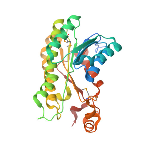
Structure of a short-chain dehydrogenase/reductase (SDR) within a genomic island from a clinical strain of Acinetobacter baumannii.

Shah, B.S.Tetu, S.G.Harrop, S.J.Paulsen, I.T.Mabbutt, B.C.

(2014) Acta Crystallogr F Struct Biol Commun 70: 1318-1323

  • DOI: https://doi.org/10.1107/S2053230X14019785
  • Primary Citation of Related Structures:  
    4IUY

  • PubMed Abstract: 

    Over 15% of the genome of an Australian clinical isolate of Acinetobacter baumannii occurs within genomic islands. An uncharacterized protein encoded within one island feature common to this and other International Clone II strains has been studied by X-ray crystallography. The 2.4 Å resolution structure of SDR-WM99c reveals it to be a new member of the classical short-chain dehydrogenase/reductase (SDR) superfamily. The enzyme contains a nucleotide-binding domain and, like many other SDRs, is tetrameric in form. The active site contains a catalytic tetrad (Asn117, Ser146, Tyr159 and Lys163) and water molecules occupying the presumed NADP cofactor-binding pocket. An adjacent cleft is capped by a relatively mobile helical subdomain, which is well positioned to control substrate access.


  • Organizational Affiliation

    Department of Chemistry and Biomolecular Sciences, Macquarie University, Research Park Drive, Sydney, NSW 2109, Australia.


Macromolecules
Find similar proteins by:  (by identity cutoff)  |  3D Structure
Entity ID: 1
MoleculeChains Sequence LengthOrganismDetailsImage
Short chain dehydrogenase/reductase274Acinetobacter baumannii ACICUMutation(s): 0 
Entity Groups  
Sequence Clusters30% Identity50% Identity70% Identity90% Identity95% Identity100% Identity
Sequence Annotations
Expand
  • Reference Sequence
Small Molecules
Modified Residues  1 Unique
IDChains TypeFormula2D DiagramParent
MSE
Query on MSE
A
B
C [auth E]
D [auth C]
E [auth H]
A,
B,
C [auth E],
D [auth C],
E [auth H],
F [auth D],
G [auth F],
H [auth G]
L-PEPTIDE LINKINGC5 H11 N O2 SeMET
Experimental Data & Validation

Experimental Data

  • Method: X-RAY DIFFRACTION
  • Resolution: 2.38 Å
  • R-Value Free: 0.203 
  • R-Value Work: 0.155 
  • R-Value Observed: 0.156 
  • Space Group: P 1 21 1
Unit Cell:
Length ( Å )Angle ( ˚ )
a = 106.219α = 90
b = 89.525β = 112.69
c = 120.905γ = 90
Software Package:
Software NamePurpose
ADSCdata collection
CCP4model building
PHENIXrefinement
XDSdata reduction
XDSdata scaling
CCP4phasing

Structure Validation

View Full Validation Report


Entry History 

Deposition Data

Revision History  (Full details and data files)

  • Version 1.0: 2013-03-06
    Type: Initial release
  • Version 1.1: 2020-04-29
    Changes: Advisory, Database references, Derived calculations
  • Version 1.2: 2023-09-20
    Changes: Advisory, Data collection, Database references, Refinement description
  • Version 1.3: 2023-12-06
    Changes: Data collection