4ISW

Crystal Structure of Phosphorylated C.elegans Thymidylate Synthase in Complex with dUMP


Experimental Data Snapshot

  • Method: X-RAY DIFFRACTION
  • Resolution: 3.14 Å
  • R-Value Free: 0.270 
  • R-Value Work: 0.234 
  • R-Value Observed: 0.236 

wwPDB Validation   3D Report Full Report


Ligand Structure Quality Assessment 


This is version 1.1 of the entry. See complete history


Literature

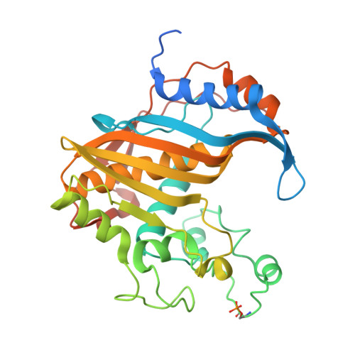
Crystal structure of phosphoramide-phosphorylated thymidylate synthase reveals pSer127, reflecting probably pHis to pSer phosphotransfer.

Wilk, P.Jarmua, A.Ruman, T.Banaszak, K.Rypniewski, W.Ciesla, J.Dowiercia, A.Rode, W.

(2013) Bioorg Chem 52C: 44-49

  • DOI: https://doi.org/10.1016/j.bioorg.2013.11.003
  • Primary Citation of Related Structures:  
    4ISW

  • PubMed Abstract: 

    Crystal structure is presented of the binary complex between potassium phosphoramidate-phosphorylated recombinant C. elegans thymidylate synthase and dUMP. On each monomer a single phosphoserine residue (Ser127) was identified, instead of expected phosphohistidine. As (31)P NMR studies of both the phosphorylated protein and of potassium phosphoramidate potential to phosphorylate different amino acids point to histidine as the only possible site of the modification, thermodynamically favored intermolecular phosphotransfer from histidine to serine is suggested.


  • Organizational Affiliation

    Nencki Institute of Experimental Biology, Polish Academy of Sciences, Warszawa, Poland.


Macromolecules
Find similar proteins by:  (by identity cutoff)  |  3D Structure
Entity ID: 1
MoleculeChains Sequence LengthOrganismDetailsImage
Thymidylate synthase
A, B
315Caenorhabditis elegansMutation(s): 0 
EC: 2.1.1.45
UniProt
Find proteins for Q9Y052 (Caenorhabditis elegans)
Explore Q9Y052 
Go to UniProtKB:  Q9Y052
Entity Groups  
Sequence Clusters30% Identity50% Identity70% Identity90% Identity95% Identity100% Identity
UniProt GroupQ9Y052
Sequence Annotations
Expand
  • Reference Sequence
Small Molecules
Ligands 1 Unique
IDChains Name / Formula / InChI Key2D Diagram3D Interactions
UMP
Query on UMP

Download Ideal Coordinates CCD File 
C [auth A],
D [auth B]
2'-DEOXYURIDINE 5'-MONOPHOSPHATE
C9 H13 N2 O8 P
JSRLJPSBLDHEIO-SHYZEUOFSA-N
Modified Residues  1 Unique
IDChains TypeFormula2D DiagramParent
SEP
Query on SEP
A, B
L-PEPTIDE LINKINGC3 H8 N O6 PSER
Experimental Data & Validation

Experimental Data

  • Method: X-RAY DIFFRACTION
  • Resolution: 3.14 Å
  • R-Value Free: 0.270 
  • R-Value Work: 0.234 
  • R-Value Observed: 0.236 
  • Space Group: P 32 2 1
Unit Cell:
Length ( Å )Angle ( ˚ )
a = 133.436α = 90
b = 133.436β = 90
c = 153.969γ = 120
Software Package:
Software NamePurpose
SCALEPACKdata scaling
PHASERphasing
REFMACrefinement
PDB_EXTRACTdata extraction

Structure Validation

View Full Validation Report


Ligand Structure Quality Assessment 


Entry History 

Deposition Data

Revision History  (Full details and data files)

  • Version 1.0: 2013-12-11
    Type: Initial release
  • Version 1.1: 2014-01-15
    Changes: Database references