4IQY

Crystal structure of the human protein-proximal ADP-ribosyl-hydrolase MacroD2


Experimental Data Snapshot

  • Method: X-RAY DIFFRACTION
  • Resolution: 1.55 Å
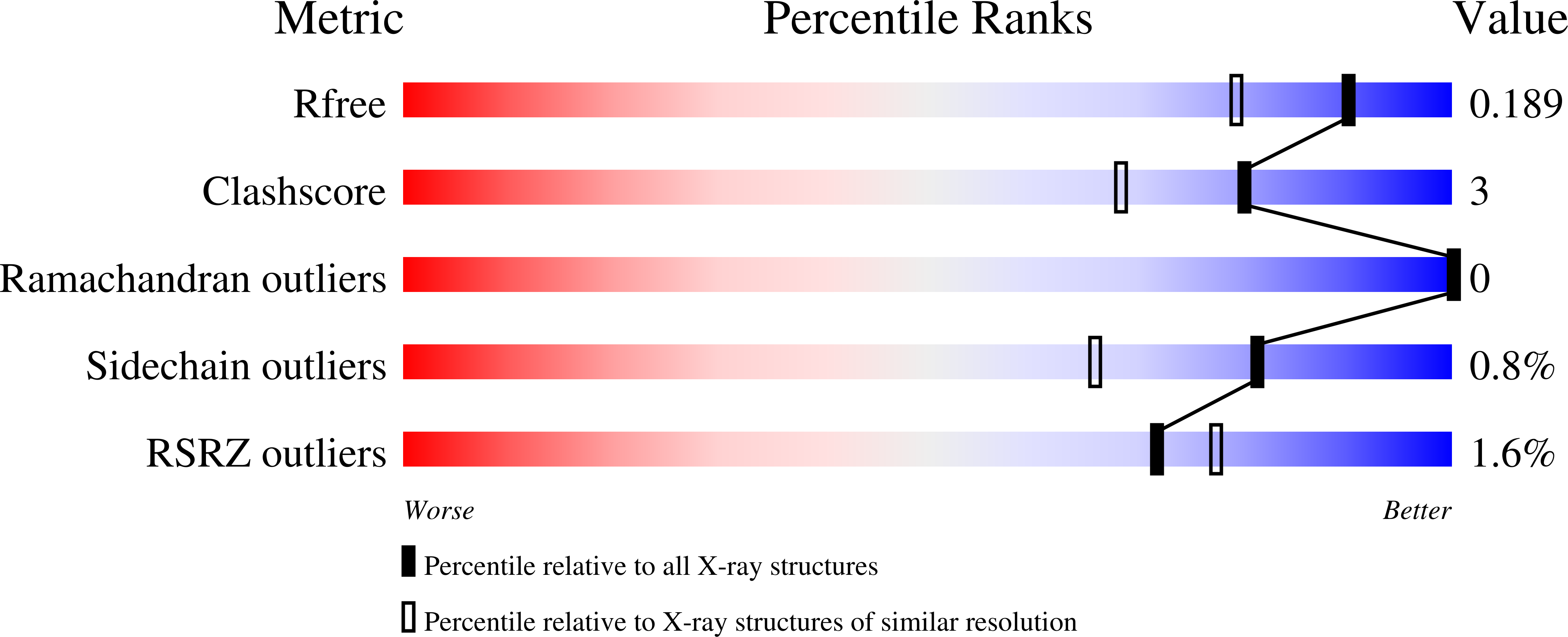
  • R-Value Free: 0.187 
  • R-Value Work: 0.158 
  • R-Value Observed: 0.160 

wwPDB Validation   3D Report Full Report


Ligand Structure Quality Assessment 


This is version 1.2 of the entry. See complete history


Literature

A family of macrodomain proteins reverses cellular mono-ADP-ribosylation.

Jankevicius, G.Hassler, M.Golia, B.Rybin, V.Zacharias, M.Timinszky, G.Ladurner, A.G.

(2013) Nat Struct Mol Biol 20: 508-514

  • DOI: https://doi.org/10.1038/nsmb.2523
  • Primary Citation of Related Structures:  
    4IQY

  • PubMed Abstract: 

    ADP-ribosylation is a reversible post-translational modification with wide-ranging biological functions in all kingdoms of life. A variety of enzymes use NAD(+) to transfer either single or multiple ADP-ribose (ADPr) moieties onto distinct amino acid substrates, often in response to DNA damage or other stresses. Poly-ADPr-glycohydrolase readily reverses poly-ADP-ribosylation induced by the DNA-damage sensor PARP1 and other enzymes, but it does not remove the most proximal ADPr linked to the target amino acid. Searches for enzymes capable of fully reversing cellular mono-ADP-ribosylation back to the unmodified state have proved elusive, which leaves a gap in the understanding of this modification. Here, we identify a family of macrodomain enzymes present in viruses, yeast and animals that reverse cellular ADP-ribosylation by acting on mono-ADP-ribosylated substrates. Our discoveries establish the complete reversibility of PARP-catalyzed cellular ADP-ribosylation as a regulatory modification.


  • Organizational Affiliation

    Butenandt Institute of Physiological Chemistry, Ludwig Maximilians University of Munich, Munich, Germany.


Macromolecules
Find similar proteins by:  (by identity cutoff)  |  3D Structure
Entity ID: 1
MoleculeChains Sequence LengthOrganismDetailsImage
O-acetyl-ADP-ribose deacetylase MACROD2
A, B
240Homo sapiensMutation(s): 0 
Gene Names: MACROD2C20orf133
EC: 3.5.1
UniProt & NIH Common Fund Data Resources
Find proteins for A1Z1Q3 (Homo sapiens)
Explore A1Z1Q3 
Go to UniProtKB:  A1Z1Q3
PHAROS:  A1Z1Q3
GTEx:  ENSG00000172264 
Entity Groups  
Sequence Clusters30% Identity50% Identity70% Identity90% Identity95% Identity100% Identity
UniProt GroupA1Z1Q3
Sequence Annotations
Expand
  • Reference Sequence
Small Molecules
Ligands 2 Unique
IDChains Name / Formula / InChI Key2D Diagram3D Interactions
AR6
Query on AR6

Download Ideal Coordinates CCD File 
C [auth A],
E [auth B]
[(2R,3S,4R,5R)-5-(6-AMINOPURIN-9-YL)-3,4-DIHYDROXY-OXOLAN-2-YL]METHYL [HYDROXY-[[(2R,3S,4R,5S)-3,4,5-TRIHYDROXYOXOLAN-2-YL]METHOXY]PHOSPHORYL] HYDROGEN PHOSPHATE
C15 H23 N5 O14 P2
SRNWOUGRCWSEMX-ZQSHOCFMSA-N
MG
Query on MG

Download Ideal Coordinates CCD File 
D [auth A],
F [auth B]
MAGNESIUM ION
Mg
JLVVSXFLKOJNIY-UHFFFAOYSA-N
Experimental Data & Validation

Experimental Data

  • Method: X-RAY DIFFRACTION
  • Resolution: 1.55 Å
  • R-Value Free: 0.187 
  • R-Value Work: 0.158 
  • R-Value Observed: 0.160 
  • Space Group: P 1
Unit Cell:
Length ( Å )Angle ( ˚ )
a = 40.713α = 69.82
b = 49.883β = 72.35
c = 66.972γ = 86.08
Software Package:
Software NamePurpose
MxCuBEdata collection
PHASERphasing
PHENIXrefinement
XDSdata reduction
SCALAdata scaling

Structure Validation

View Full Validation Report


Ligand Structure Quality Assessment 


Entry History 

Deposition Data

Revision History  (Full details and data files)

  • Version 1.0: 2013-03-06
    Type: Initial release
  • Version 1.1: 2013-04-17
    Changes: Database references
  • Version 1.2: 2023-09-20
    Changes: Data collection, Database references, Derived calculations, Refinement description, Structure summary