4IQR

Multi-Domain Organization of the HNF4alpha Nuclear Receptor Complex on DNA


Experimental Data Snapshot

  • Method: X-RAY DIFFRACTION
  • Resolution: 2.90 Å
  • R-Value Free: 0.278 
  • R-Value Work: 0.239 
  • R-Value Observed: 0.241 

wwPDB Validation   3D Report Full Report


Ligand Structure Quality Assessment 


This is version 1.2 of the entry. See complete history


Literature

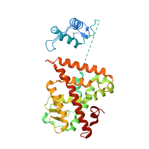
Multidomain integration in the structure of the HNF-4 alpha nuclear receptor complex.

Chandra, V.Huang, P.Potluri, N.Wu, D.Kim, Y.Rastinejad, F.

(2013) Nature 495: 394-398

  • DOI: https://doi.org/10.1038/nature11966
  • Primary Citation of Related Structures:  
    4IQR

  • PubMed Abstract: 

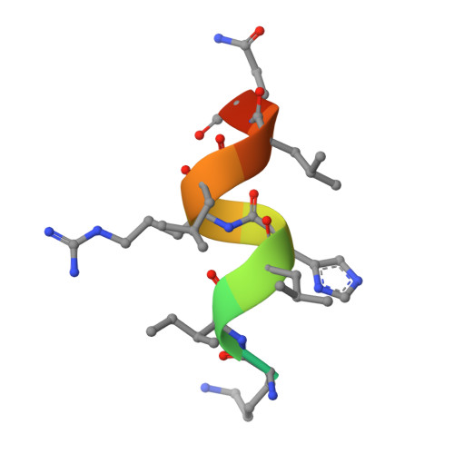
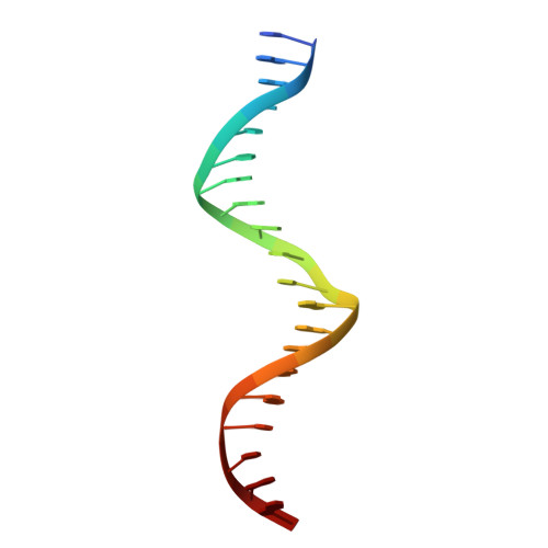
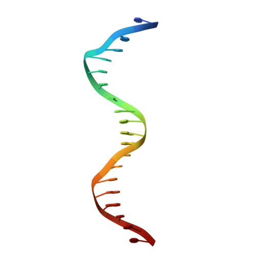
    The hepatocyte nuclear factor 4α (HNF-4α; also known as NR2A1) is a member of the nuclear receptor (NR) family of transcription factors, which have conserved DNA-binding domains and ligand-binding domains. HNF-4α is the most abundant DNA-binding protein in the liver, where some 40% of the actively transcribed genes have a HNF-4α response element. These regulated genes are largely involved in the hepatic gluconeogenic program and lipid metabolism. In the pancreas HNF-4α is also a master regulator, controlling an estimated 11% of islet genes. HNF-4α protein mutations are linked to maturity-onset diabetes of the young, type 1 (MODY1) and hyperinsulinaemic hypoglycaemia. Previous structural analyses of NRs, although productive in elucidating the structure of individual domains, have lagged behind in revealing the connectivity patterns of NR domains. Here we describe the 2.9 Å crystal structure of the multidomain human HNF-4α homodimer bound to its DNA response element and coactivator-derived peptides. A convergence zone connects multiple receptor domains in an asymmetric fashion, joining distinct elements from each monomer. An arginine target of PRMT1 methylation protrudes directly into this convergence zone and sustains its integrity. A serine target of protein kinase C is also responsible for maintaining domain-domain interactions. These post-translational modifications lead to changes in DNA binding by communicating through the tightly connected surfaces of the quaternary fold. We find that some MODY1 mutations, positioned on the ligand-binding domain and hinge regions of the receptor, compromise DNA binding at a distance by communicating through the interjunctional surfaces of the complex. The overall domain representation of the HNF-4α homodimer is different from that of the PPAR-γ-RXR-α heterodimer, even when both NR complexes are assembled on the same DNA element. Our findings suggest that unique quaternary folds and interdomain connections in NRs could be exploited by small-molecule allosteric modulators that affect distal functions in these polypeptides.


  • Organizational Affiliation

    Metabolic Signaling and Disease Program, Sanford-Burnham Medical Research Institute, Orlando, Florida 32827, USA.


Macromolecules

Find similar proteins by:  (by identity cutoff)  |  3D Structure
Entity ID: 1
MoleculeChains Sequence LengthOrganismDetailsImage
Hepatocyte nuclear factor 4-alpha
A, B, E, F
338Homo sapiensMutation(s): 0 
Gene Names: HNF4AHNF4NR2A1TCF14
UniProt & NIH Common Fund Data Resources
Find proteins for P41235 (Homo sapiens)
Explore P41235 
Go to UniProtKB:  P41235
PHAROS:  P41235
GTEx:  ENSG00000101076 
Entity Groups  
Sequence Clusters30% Identity50% Identity70% Identity90% Identity95% Identity100% Identity
UniProt GroupP41235
Sequence Annotations
Expand
  • Reference Sequence

Find similar proteins by:  Sequence   |   3D Structure  

Entity ID: 4
MoleculeChains Sequence LengthOrganismDetailsImage
Nuclear receptor coactivator 2
I, J, K, L
13Homo sapiensMutation(s): 0 
UniProt & NIH Common Fund Data Resources
Find proteins for Q15596 (Homo sapiens)
Explore Q15596 
Go to UniProtKB:  Q15596
PHAROS:  Q15596
GTEx:  ENSG00000140396 
Entity Groups  
Sequence Clusters30% Identity50% Identity70% Identity90% Identity95% Identity100% Identity
UniProt GroupQ15596
Sequence Annotations
Expand
  • Reference Sequence

Find similar nucleic acids by:  Sequence   |   3D Structure  

Entity ID: 2
MoleculeChains LengthOrganismImage
DNA (5'-D(*GP*GP*AP*AP*CP*TP*AP*GP*GP*TP*CP*AP*AP*AP*GP*GP*TP*CP*AP*G)-3')
C, G
20N/A
Sequence Annotations
Expand
  • Reference Sequence

Find similar nucleic acids by:  Sequence   |   3D Structure  

Entity ID: 3
MoleculeChains LengthOrganismImage
DNA (5'-D(*CP*CP*TP*GP*AP*CP*CP*TP*TP*TP*GP*AP*CP*CP*TP*AP*GP*TP*TP*C)-3')
D, H
20N/A
Sequence Annotations
Expand
  • Reference Sequence
Small Molecules
Ligands 2 Unique
IDChains Name / Formula / InChI Key2D Diagram3D Interactions
MYR
Query on MYR

Download Ideal Coordinates CCD File 
M [auth A],
P [auth B],
S [auth E],
V [auth F]
MYRISTIC ACID
C14 H28 O2
TUNFSRHWOTWDNC-UHFFFAOYSA-N
ZN
Query on ZN

Download Ideal Coordinates CCD File 
N [auth A]
O [auth A]
Q [auth B]
R [auth B]
T [auth E]
N [auth A],
O [auth A],
Q [auth B],
R [auth B],
T [auth E],
U [auth E],
W [auth F],
X [auth F]
ZINC ION
Zn
PTFCDOFLOPIGGS-UHFFFAOYSA-N
Experimental Data & Validation

Experimental Data

  • Method: X-RAY DIFFRACTION
  • Resolution: 2.90 Å
  • R-Value Free: 0.278 
  • R-Value Work: 0.239 
  • R-Value Observed: 0.241 
  • Space Group: P 1 21 1
Unit Cell:
Length ( Å )Angle ( ˚ )
a = 119.786α = 90
b = 57.596β = 96.3
c = 166.305γ = 90
Software Package:
Software NamePurpose
HKL-3000data collection
PHASERphasing
CNSrefinement
HKL-3000data reduction
HKL-3000data scaling

Structure Validation

View Full Validation Report


Ligand Structure Quality Assessment 


Entry History 

Deposition Data

Revision History  (Full details and data files)

  • Version 1.0: 2013-03-20
    Type: Initial release
  • Version 1.1: 2013-10-23
    Changes: Database references
  • Version 1.2: 2023-09-20
    Changes: Data collection, Database references, Derived calculations, Refinement description