Structural and dynamic insights into substrate binding and catalysis of human lipocalin prostaglandin D synthase.
Lim, S.M., Chen, D., Teo, H., Roos, A., Jansson, A.E., Nyman, T., Tresaugues, L., Pervushin, K., Nordlund, P.(2013) J Lipid Res 54: 1630-1643
- PubMed: 23526831
- DOI: https://doi.org/10.1194/jlr.M035410
- Primary Citation of Related Structures:
2WWP, 4IMN, 4IMO - PubMed Abstract:
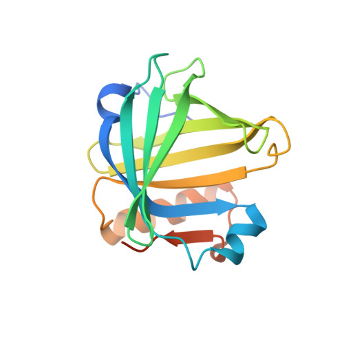
Lipocalin prostaglandin D synthase (L-PGDS) regulates synthesis of an important inflammatory and signaling mediator, prostaglandin D2 (PGD2). Here, we used structural, biophysical, and biochemical approaches to address the mechanistic aspects of substrate entry, catalysis, and product exit of this enzyme. Structure of human L-PGDS was solved in a complex with a substrate analog (SA) and in ligand-free form. Its catalytic Cys 65 thiol group was found in two different conformations, each making a distinct hydrogen bond network to neighboring residues. These help in elucidating the mechanism of the cysteine nucleophile activation. Electron density for ligand observed in the active site defined the substrate binding regions, but did not allow unambiguous fitting of the SA. To further understand ligand binding, we used NMR spectroscopy to map the binding sites and to show the dynamics of protein-substrate and protein-product interactions. A model for ligand binding at the catalytic site is proposed, showing a second binding site involved in ligand exit and entry. NMR chemical shift perturbations and NMR resonance line-width alterations (observed as changes of intensity in two-dimensional cross-peaks in [¹H,¹⁵N]-transfer relaxation optimization spectroscopy) for residues at the Ω loop (A-B loop), E-F loop, and G-H loop besides the catalytic sites indicate involvement of these residues in ligand entry/egress.
Organizational Affiliation:
Division of Structural Biology and Biochemistry, Nanyang Technological University, Singapore; and; Department of Medical Biochemistry and Biophysics, Karolinska Institutet, Stockholm, Sweden.