


2.39 Angstrom X-ray Crystal structure of human ACMSD
Liu, F., Liu, A.To be published.
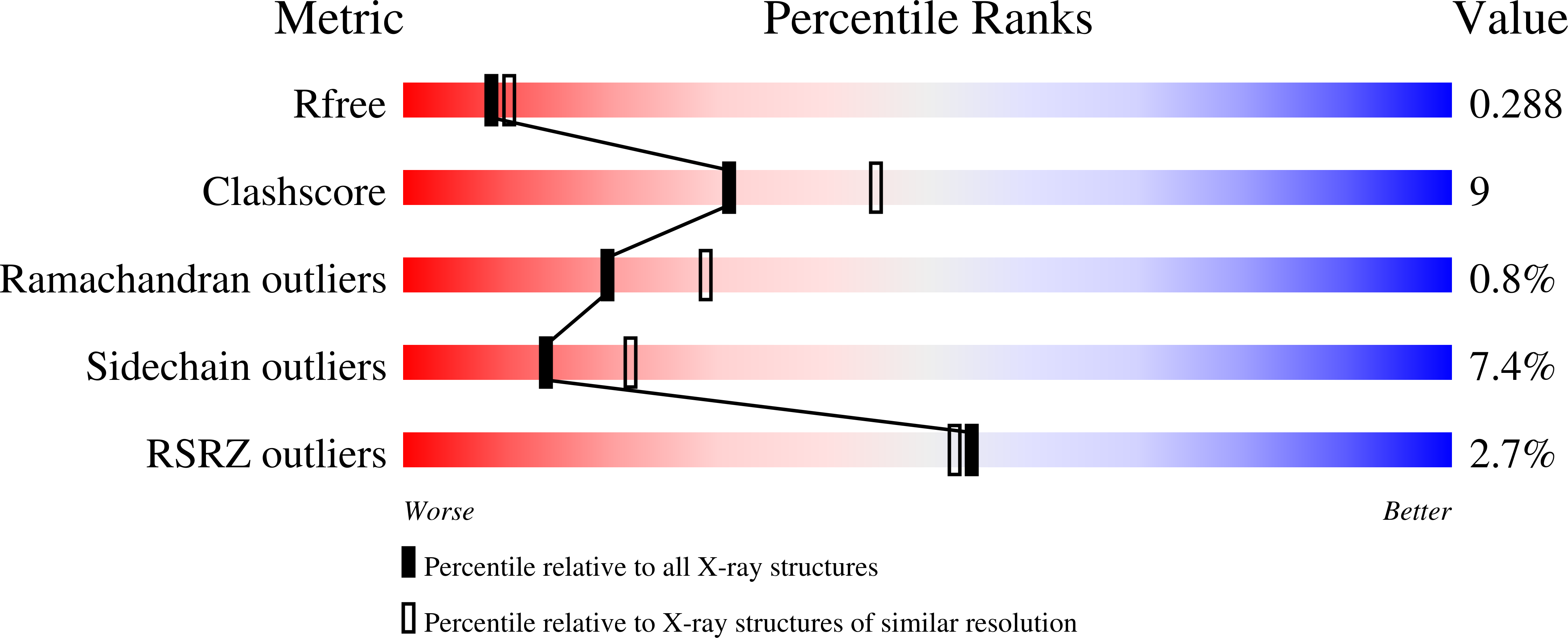
Experimental Data Snapshot
wwPDB Validation 3D Report Full Report
Entity ID: 1 | |||||
|---|---|---|---|---|---|
| Molecule | Chains | Sequence Length | Organism | Details | Image |
| 2-amino-3-carboxymuconate-6-semialdehyde decarboxylase | 332 | Homo sapiens | Mutation(s): 0 Gene Names: ACMSD, human EC: 4.1.1.45 | 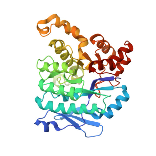 | |
UniProt & NIH Common Fund Data Resources | |||||
Find proteins for Q8TDX5 (Homo sapiens) Explore Q8TDX5 Go to UniProtKB: Q8TDX5 | |||||
PHAROS: Q8TDX5 GTEx: ENSG00000153086 | |||||
Entity Groups | |||||
| Sequence Clusters | 30% Identity50% Identity70% Identity90% Identity95% Identity100% Identity | ||||
| UniProt Group | Q8TDX5 | ||||
Sequence AnnotationsExpand | |||||
| |||||
| Ligands 1 Unique | |||||
|---|---|---|---|---|---|
| ID | Chains | Name / Formula / InChI Key | 2D Diagram | 3D Interactions | |
| ZN Query on ZN | G [auth A] H [auth B] I [auth C] J [auth D] K [auth E] | ZINC ION Zn PTFCDOFLOPIGGS-UHFFFAOYSA-N |  | ||
| Length ( Å ) | Angle ( ˚ ) |
|---|---|
| a = 89.112 | α = 90 |
| b = 101.878 | β = 90 |
| c = 233.455 | γ = 90 |
| Software Name | Purpose |
|---|---|
| REFMAC | refinement |
| PDB_EXTRACT | data extraction |
| SERGUI | data collection |
| HKL-2000 | data reduction |
| HKL-2000 | data scaling |
| MOLREP | phasing |
RCSB PDB (citation) is hosted by
RCSB PDB is a member of the

