4I9O

Crystal Structure of GACKIX L664C Tethered to 1-10


Experimental Data Snapshot

  • Method: X-RAY DIFFRACTION
  • Resolution: 2.00 Å
  • R-Value Free: 0.233 
  • R-Value Work: 0.206 
  • R-Value Observed: 0.208 

wwPDB Validation   3D Report Full Report


Ligand Structure Quality Assessment 


This is version 1.2 of the entry. See complete history


Literature

Ordering a dynamic protein via a small-molecule stabilizer.

Wang, N.Majmudar, C.Y.Pomerantz, W.C.Gagnon, J.K.Sadowsky, J.D.Meagher, J.L.Johnson, T.K.Stuckey, J.A.Brooks, C.L.Wells, J.A.Mapp, A.K.

(2013) J Am Chem Soc 135: 3363-3366

  • DOI: https://doi.org/10.1021/ja3122334
  • Primary Citation of Related Structures:  
    4I9O

  • PubMed Abstract: 

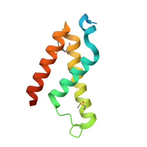
    Like many coactivators, the GACKIX domain of the master coactivator CBP/p300 recognizes transcriptional activators of diverse sequence composition via dynamic binding surfaces. The conformational dynamics of GACKIX that underlie its function also render it especially challenging for structural characterization. We have found that the ligand discovery strategy of Tethering is an effective method for identifying small-molecule fragments that stabilize the GACKIX domain, enabling for the first time the crystallographic characterization of this important motif. The 2.0 Å resolution structure of GACKIX complexed to a small molecule was further analyzed by molecular dynamics simulations, which revealed the importance of specific side-chain motions that remodel the activator binding site in order to accommodate binding partners of distinct sequence and size. More broadly, these results suggest that Tethering can be a powerful strategy for identifying small-molecule stabilizers of conformationally malleable proteins, thus facilitating their structural characterization and accelerating the discovery of small-molecule modulators.


  • Organizational Affiliation

    Program in Chemical Biology, University of Michigan, Ann Arbor, Michigan 48109, United States.


Macromolecules
Find similar proteins by:  (by identity cutoff)  |  3D Structure
Entity ID: 1
MoleculeChains Sequence LengthOrganismDetailsImage
CREB-binding protein101Mus musculusMutation(s): 1 
Gene Names: CrebbpCbp
EC: 2.3.1.48
UniProt
Find proteins for P45481 (Mus musculus)
Explore P45481 
Go to UniProtKB:  P45481
Entity Groups  
Sequence Clusters30% Identity50% Identity70% Identity90% Identity95% Identity100% Identity
UniProt GroupP45481
Sequence Annotations
Expand
  • Reference Sequence
Small Molecules
Ligands 2 Unique
IDChains Name / Formula / InChI Key2D Diagram3D Interactions
KI1
Query on KI1

Download Ideal Coordinates CCD File 
B [auth A]1-{4-[4-chloro-3-(trifluoromethyl)phenyl]-4-hydroxypiperidin-1-yl}-3-sulfanylpropan-1-one
C15 H17 Cl F3 N O2 S
STKPEUISNQZVRV-UHFFFAOYSA-N
EDO
Query on EDO

Download Ideal Coordinates CCD File 
C [auth A],
D [auth A]
1,2-ETHANEDIOL
C2 H6 O2
LYCAIKOWRPUZTN-UHFFFAOYSA-N
Modified Residues  1 Unique
IDChains TypeFormula2D DiagramParent
MSE
Query on MSE
A
L-PEPTIDE LINKINGC5 H11 N O2 SeMET
Binding Affinity Annotations 
IDSourceBinding Affinity
KI1 PDBBind:  4I9O Kd: 1900 (nM) from 1 assay(s)
Experimental Data & Validation

Experimental Data

  • Method: X-RAY DIFFRACTION
  • Resolution: 2.00 Å
  • R-Value Free: 0.233 
  • R-Value Work: 0.206 
  • R-Value Observed: 0.208 
  • Space Group: P 43 21 2
Unit Cell:
Length ( Å )Angle ( ˚ )
a = 48.33α = 90
b = 48.33β = 90
c = 85.464γ = 90
Software Package:
Software NamePurpose
DENZOdata reduction
SCALEPACKdata scaling
BUSTER-TNTrefinement
PDB_EXTRACTdata extraction
MD2data collection
HKL-2000data reduction
HKL-2000data scaling
PHENIXphasing
BUSTERrefinement

Structure Validation

View Full Validation Report


Ligand Structure Quality Assessment 


Entry History 

Deposition Data

Revision History  (Full details and data files)

  • Version 1.0: 2013-03-06
    Type: Initial release
  • Version 1.1: 2013-04-17
    Changes: Database references
  • Version 1.2: 2017-11-15
    Changes: Advisory, Refinement description