4I0K

Crystal structure of murine B7-H3 extracellular domain


Experimental Data Snapshot

  • Method: X-RAY DIFFRACTION
  • Resolution: 2.97 Å
  • R-Value Free: 0.250 
  • R-Value Work: 0.215 
  • R-Value Observed: 0.217 

wwPDB Validation   3D Report Full Report


Ligand Structure Quality Assessment 


This is version 2.1 of the entry. See complete history


Literature

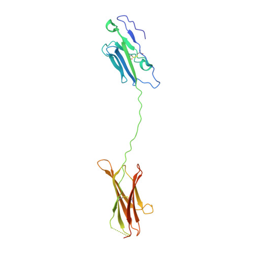
Structure and T cell inhibition properties of B7 family member, B7-H3.

Vigdorovich, V.Ramagopal, U.A.Lazar-Molnar, E.Sylvestre, E.Lee, J.S.Hofmeyer, K.A.Zang, X.Nathenson, S.G.Almo, S.C.

(2013) Structure 21: 707-717

  • DOI: https://doi.org/10.1016/j.str.2013.03.003
  • Primary Citation of Related Structures:  
    4I0K

  • PubMed Abstract: 

    T cell activity is controlled by a combination of antigen-dependent signaling through the T cell receptor and a set of auxiliary signals delivered through antigen-independent interactions, including the recognition of the B7 family of ligands. B7-H3 is a recently identified B7 family member that is strongly overexpressed in a range of cancers and correlates with poor prognosis. We report the crystal structure of murine B7-H3 at a 3 Å resolution, which provides a model for the organization of the IgV and IgC domains within the ectodomain. We demonstrate that B7-H3 inhibits T cell proliferation and show that the FG loop of the IgV domain plays a critical role in this function. B7-H3 crystallized as an unusual dimer arising from the exchange of the G strands in the IgV domains of partner molecules. This arrangement, in combination with previous reports, highlights the dynamic nature and plasticity of the immunoglobulin fold.


  • Organizational Affiliation

    Department of Microbiology and Immunology, Albert Einstein College of Medicine, Bronx, NY 10461, USA. vladimir.vigdorovich@gmail.com


Macromolecules
Find similar proteins by:  (by identity cutoff)  |  3D Structure
Entity ID: 1
MoleculeChains Sequence LengthOrganismDetailsImage
CD276 antigen222Mus musculusMutation(s): 0 
Gene Names: B7-H3B7h3Cd276
UniProt & NIH Common Fund Data Resources
Find proteins for Q8VE98 (Mus musculus)
Explore Q8VE98 
Go to UniProtKB:  Q8VE98
IMPC:  MGI:2183926
Entity Groups  
Sequence Clusters30% Identity50% Identity70% Identity90% Identity95% Identity100% Identity
UniProt GroupQ8VE98
Sequence Annotations
Expand
  • Reference Sequence
Oligosaccharides

Help

Entity ID: 2
MoleculeChains Length2D Diagram Glycosylation3D Interactions
alpha-D-mannopyranose-(1-6)-beta-D-mannopyranose-(1-4)-2-acetamido-2-deoxy-beta-D-glucopyranose-(1-4)-2-acetamido-2-deoxy-beta-D-glucopyranose
B
4N-Glycosylation
Glycosylation Resources
GlyTouCan:  G22573RC
GlyCosmos:  G22573RC
GlyGen:  G22573RC
Experimental Data & Validation

Experimental Data

  • Method: X-RAY DIFFRACTION
  • Resolution: 2.97 Å
  • R-Value Free: 0.250 
  • R-Value Work: 0.215 
  • R-Value Observed: 0.217 
  • Space Group: P 61 2 2
Unit Cell:
Length ( Å )Angle ( ˚ )
a = 100.92α = 90
b = 100.92β = 90
c = 188.18γ = 120
Software Package:
Software NamePurpose
SCALAdata scaling
REFMACrefinement
PDB_EXTRACTdata extraction
CBASSdata collection
MOSFLMdata reduction
BALBESphasing

Structure Validation

View Full Validation Report


Ligand Structure Quality Assessment 


Entry History 

Revision History  (Full details and data files)

  • Version 1.0: 2012-12-05
    Type: Initial release
  • Version 1.1: 2016-11-16
    Changes: Database references
  • Version 2.0: 2020-07-29
    Type: Remediation
    Reason: Carbohydrate remediation
    Changes: Advisory, Atomic model, Data collection, Database references, Derived calculations, Structure summary
  • Version 2.1: 2023-09-20
    Changes: Data collection, Database references, Refinement description, Structure summary