4HXY

PlmKR1-Ketoreductase from the first module of phoslactomycin biosynthesis in Streptomyces sp. HK803


Experimental Data Snapshot

  • Method: X-RAY DIFFRACTION
  • Resolution: 1.68 Å
  • R-Value Free: 0.188 
  • R-Value Work: 0.155 
  • R-Value Observed: 0.156 

wwPDB Validation   3D Report Full Report


Ligand Structure Quality Assessment 


This is version 2.0 of the entry. See complete history


Literature

Structural and Stereochemical Analysis of a Modular Polyketide Synthase Ketoreductase Domain Required for the Generation of a cis-Alkene.

Bonnett, S.A.Whicher, J.R.Papireddy, K.Florova, G.Smith, J.L.Reynolds, K.A.

(2013) Chem Biol 20: 772-783

  • DOI: https://doi.org/10.1016/j.chembiol.2013.04.014
  • Primary Citation of Related Structures:  
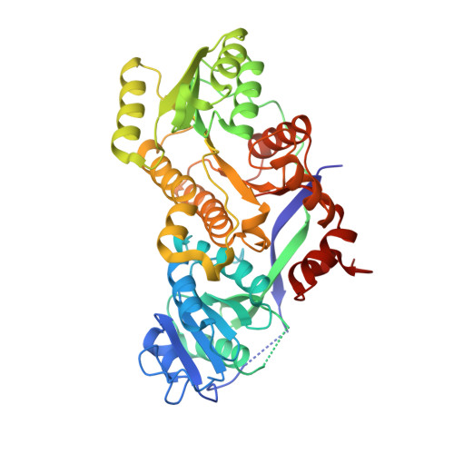
    4HXY

  • PubMed Abstract: 

    The formation of an activated cis-3-cyclohexylpropenoic acid by Plm1, the first extension module of the phoslactomycin polyketide synthase, is proposed to occur through an L-3-hydroxyacyl-intermediate as a result of ketoreduction by an A-type ketoreductase (KR). Here, we demonstrate that the KR domain of Plm1 (PlmKR1) catalyzes the formation of an L-3-hydroxyacyl product. The crystal structure of PlmKR1 revealed a well-ordered active site with a nearby Trp residue characteristic of A-type KRs. Structural comparison of PlmKR1 with B-type KRs that produce D-3-hydroxyacyl intermediates revealed significant differences. The active site of cofactor-bound A-type KRs is in a catalysis-ready state, whereas cofactor-bound B-type KRs are in a precatalytic state. Furthermore, the closed lid loop in substrate-bound A-type KRs restricts active site access from all but one direction, which is proposed to control the stereochemistry of ketoreduction.


  • Organizational Affiliation

    Department of Chemistry, Portland State University, Portland, OR. 97201.


Macromolecules
Find similar proteins by:  (by identity cutoff)  |  3D Structure
Entity ID: 1
MoleculeChains Sequence LengthOrganismDetailsImage
Plm1
A, B
438Streptomyces sp. HK803Mutation(s): 0 
Gene Names: plm1
UniProt
Find proteins for Q6V1M8 (Streptomyces sp. HK803)
Explore Q6V1M8 
Go to UniProtKB:  Q6V1M8
Entity Groups  
Sequence Clusters30% Identity50% Identity70% Identity90% Identity95% Identity100% Identity
UniProt GroupQ6V1M8
Sequence Annotations
Expand
  • Reference Sequence
Small Molecules
Ligands 2 Unique
IDChains Name / Formula / InChI Key2D Diagram3D Interactions
NDP
Query on NDP

Download Ideal Coordinates CCD File 
C [auth A],
D [auth B]
NADPH DIHYDRO-NICOTINAMIDE-ADENINE-DINUCLEOTIDE PHOSPHATE
C21 H30 N7 O17 P3
ACFIXJIJDZMPPO-NNYOXOHSSA-N
ACA
Query on ACA

Download Ideal Coordinates CCD File 
E [auth B]6-AMINOHEXANOIC ACID
C6 H13 N O2
SLXKOJJOQWFEFD-UHFFFAOYSA-N
Experimental Data & Validation

Experimental Data

  • Method: X-RAY DIFFRACTION
  • Resolution: 1.68 Å
  • R-Value Free: 0.188 
  • R-Value Work: 0.155 
  • R-Value Observed: 0.156 
  • Space Group: P 1 21 1
Unit Cell:
Length ( Å )Angle ( ˚ )
a = 42.157α = 90
b = 123.398β = 90.27
c = 79.884γ = 90
Software Package:
Software NamePurpose
Blu-Icedata collection
PHASERphasing
REFMACrefinement
HKL-2000data reduction
HKL-2000data scaling

Structure Validation

View Full Validation Report


Ligand Structure Quality Assessment 


Entry History 

Deposition Data

Revision History  (Full details and data files)

  • Version 1.0: 2013-07-10
    Type: Initial release
  • Version 2.0: 2023-11-15
    Changes: Atomic model, Data collection, Database references, Derived calculations