4HXI

Crystal structure of KLHL3/Cul3 complex


Experimental Data Snapshot

  • Method: X-RAY DIFFRACTION
  • Resolution: 3.51 Å
  • R-Value Free: 0.278 
  • R-Value Work: 0.244 
  • R-Value Observed: 0.248 

wwPDB Validation   3D Report Full Report


This is version 1.1 of the entry. See complete history


Literature

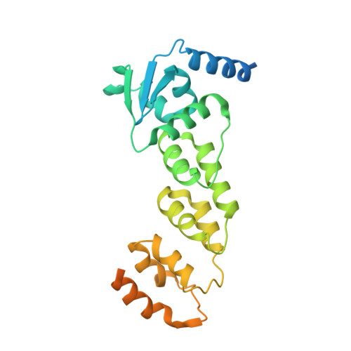
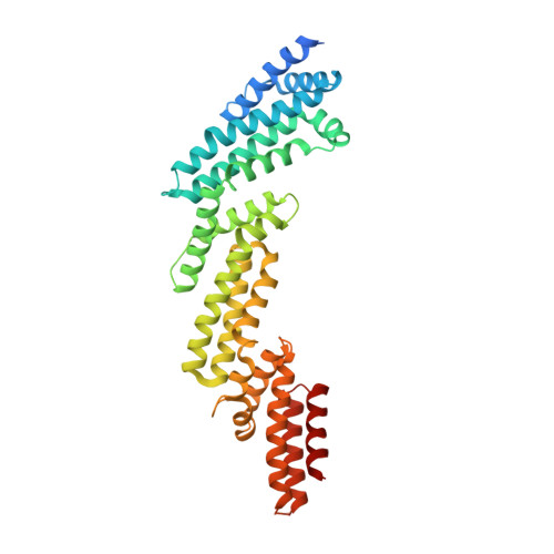
Crystal structure of KLHL3 in complex with Cullin3.

Ji, A.X.Prive, G.G.

(2013) PLoS One 8: e60445-e60445

  • DOI: https://doi.org/10.1371/journal.pone.0060445
  • Primary Citation of Related Structures:  
    4HXI

  • PubMed Abstract: 

    KLHL3 is a BTB-BACK-Kelch family protein that serves as a substrate adapter in Cullin3 (Cul3) E3 ubiquitin ligase complexes. KLHL3 is highly expressed in distal nephron tubules where it is involved in the regulation of electrolyte homeostasis and blood pressure. Mutations in KLHL3 have been identified in patients with inherited hypertension disorders, and several of the disease-associated mutations are located in the presumed Cul3 binding region. Here, we report the crystal structure of a complex between the KLHL3 BTB-BACK domain dimer and two copies of an N terminal fragment of Cul3. We use isothermal titration calorimetry to directly demonstrate that several of the disease mutations in the KLHL3 BTB-BACK domains disrupt the association with Cul3. Both the BTB and BACK domains contribute to the Cul3 interaction surface, and an extended model of the dimeric CRL3 complex places the two E2 binding sites in a suprafacial arrangement with respect to the presumed substrate-binding sites.


  • Organizational Affiliation

    Department of Biochemistry, University of Toronto, Toronto, Ontario, Canada.


Macromolecules
Find similar proteins by:  (by identity cutoff)  |  3D Structure
Entity ID: 1
MoleculeChains Sequence LengthOrganismDetailsImage
Kelch-like protein 3277Homo sapiensMutation(s): 3 
Gene Names: KLHL3KIAA1129
UniProt & NIH Common Fund Data Resources
Find proteins for Q9UH77 (Homo sapiens)
Explore Q9UH77 
Go to UniProtKB:  Q9UH77
PHAROS:  Q9UH77
GTEx:  ENSG00000146021 
Entity Groups  
Sequence Clusters30% Identity50% Identity70% Identity90% Identity95% Identity100% Identity
UniProt GroupQ9UH77
Sequence Annotations
Expand
  • Reference Sequence
Find similar proteins by:  (by identity cutoff)  |  3D Structure
Entity ID: 2
MoleculeChains Sequence LengthOrganismDetailsImage
Cullin-3386Homo sapiensMutation(s): 3 
Gene Names: CUL3KIAA0617
UniProt & NIH Common Fund Data Resources
Find proteins for Q13618 (Homo sapiens)
Explore Q13618 
Go to UniProtKB:  Q13618
PHAROS:  Q13618
GTEx:  ENSG00000036257 
Entity Groups  
Sequence Clusters30% Identity50% Identity70% Identity90% Identity95% Identity100% Identity
UniProt GroupQ13618
Sequence Annotations
Expand
  • Reference Sequence
Experimental Data & Validation

Experimental Data

  • Method: X-RAY DIFFRACTION
  • Resolution: 3.51 Å
  • R-Value Free: 0.278 
  • R-Value Work: 0.244 
  • R-Value Observed: 0.248 
  • Space Group: C 2 2 21
Unit Cell:
Length ( Å )Angle ( ˚ )
a = 40.77α = 90
b = 228.677β = 90
c = 239.989γ = 90
Software Package:
Software NamePurpose
ADSCdata collection
PHENIXmodel building
PHENIXrefinement
HKL-3000data reduction
HKL-3000data scaling
PHENIXphasing

Structure Validation

View Full Validation Report


Entry History 

Deposition Data

Revision History  (Full details and data files)

  • Version 1.0: 2013-03-13
    Type: Initial release
  • Version 1.1: 2023-11-15
    Changes: Data collection, Database references