


X-ray crystal structure of salicylic acid bound 3-hydroxyanthranilate-3,4-dioxygenase from cupriavidus metallidurans
Liu, F., Chen, L., Liu, A.To be published.
Experimental Data Snapshot
wwPDB Validation 3D Report Full Report

Entity ID: 1 | |||||
|---|---|---|---|---|---|
| Molecule | Chains | Sequence Length | Organism | Details | Image |
| 3-hydroxyanthranilate 3,4-dioxygenase | 174 | Cupriavidus metallidurans CH34 | Mutation(s): 0 Gene Names: Cupriavidus metallidurans, nbaC, Rmet_5193 EC: 1.13.11.6 | 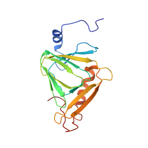 | |
UniProt | |||||
Find proteins for Q1LCS4 (Cupriavidus metallidurans (strain ATCC 43123 / DSM 2839 / NBRC 102507 / CH34)) Explore Q1LCS4 Go to UniProtKB: Q1LCS4 | |||||
Entity Groups | |||||
| Sequence Clusters | 30% Identity50% Identity70% Identity90% Identity95% Identity100% Identity | ||||
| UniProt Group | Q1LCS4 | ||||
Sequence AnnotationsExpand | |||||
| |||||
| Ligands 2 Unique | |||||
|---|---|---|---|---|---|
| ID | Chains | Name / Formula / InChI Key | 2D Diagram | 3D Interactions | |
| SAL Query on SAL | D [auth A] | 2-HYDROXYBENZOIC ACID C7 H6 O3 YGSDEFSMJLZEOE-UHFFFAOYSA-N |  | ||
| FE Query on FE | B [auth A], C [auth A] | FE (III) ION Fe VTLYFUHAOXGGBS-UHFFFAOYSA-N |  | ||
| Length ( Å ) | Angle ( ˚ ) |
|---|---|
| a = 58.561 | α = 90 |
| b = 58.561 | β = 90 |
| c = 230.99 | γ = 120 |
| Software Name | Purpose |
|---|---|
| REFMAC | refinement |
| PDB_EXTRACT | data extraction |
| SERGUI | data collection |
| DENZO | data reduction |
| SCALEPACK | data scaling |
| HKL-2000 | data scaling |
| MOLREP | phasing |
RCSB PDB (citation) is hosted by
RCSB PDB is a member of the

