4HUP

Structure of ricin A chain bound with N-(N-(pterin-7-yl)carbonylglycyl)-L-phenylalanyl)-L-phenylalanine


Experimental Data Snapshot

  • Method: X-RAY DIFFRACTION
  • Resolution: 1.70 Å
  • R-Value Free: 0.236 
  • R-Value Work: 0.209 
  • R-Value Observed: 0.211 

wwPDB Validation   3D Report Full Report


Ligand Structure Quality Assessment 


This is version 1.5 of the entry. See complete history


Literature

Peptide-conjugated pterins as inhibitors of ricin toxin A.

Saito, R.Pruet, J.M.Manzano, L.A.Jasheway, K.Monzingo, A.F.Wiget, P.A.Kamat, I.Anslyn, E.V.Robertus, J.D.

(2013) J Med Chem 56: 320-329

  • DOI: https://doi.org/10.1021/jm3016393
  • Primary Citation of Related Structures:  
    4HUO, 4HUP, 4HV3, 4HV7

  • PubMed Abstract: 

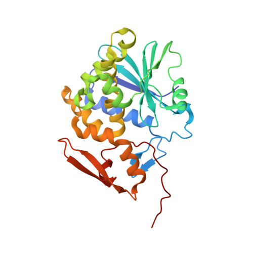
    Several 7-peptide-substituted pterins were synthesized and tested as competitive active-site inhibitors of ricin toxin A (RTA). Focus began on dipeptide conjugates, and these results further guided the construction of several tripeptide conjugates. The binding of these compounds to RTA was studied via a luminescence-based kinetic assay, as well as through X-ray crystallography. Despite the relatively polar, solvent exposed active site, several hydrophobic interactions, most commonly π-interactions not predicted by modeling programs, were identified in all of the best-performing inhibitors. Nearly all of these compounds provide IC₅₀ values in the low micromolar range.


  • Organizational Affiliation

    Department of Chemistry, Toho University, 2-2-1 Miyama, Funabashi 274-8510, Japan. anslyn@austin.utexas.edu


Macromolecules
Find similar proteins by:  (by identity cutoff)  |  3D Structure
Entity ID: 1
MoleculeChains Sequence LengthOrganismDetailsImage
RicinA [auth X]267Ricinus communisMutation(s): 0 
EC: 3.2.2.22
UniProt
Find proteins for P02879 (Ricinus communis)
Explore P02879 
Go to UniProtKB:  P02879
Entity Groups  
Sequence Clusters30% Identity50% Identity70% Identity90% Identity95% Identity100% Identity
UniProt GroupP02879
Sequence Annotations
Expand
  • Reference Sequence
Small Molecules
Binding Affinity Annotations 
IDSourceBinding Affinity
19M Binding MOAD:  4HUP IC50: 1.50e+4 (nM) from 1 assay(s)
PDBBind:  4HUP IC50: 1.50e+4 (nM) from 1 assay(s)
Experimental Data & Validation

Experimental Data

  • Method: X-RAY DIFFRACTION
  • Resolution: 1.70 Å
  • R-Value Free: 0.236 
  • R-Value Work: 0.209 
  • R-Value Observed: 0.211 
  • Space Group: P 41 21 2
Unit Cell:
Length ( Å )Angle ( ˚ )
a = 67.723α = 90
b = 67.723β = 90
c = 140.592γ = 90
Software Package:
Software NamePurpose
DENZOdata reduction
SCALEPACKdata scaling
PHENIXrefinement
PDB_EXTRACTdata extraction
StructureStudiodata collection
HKL-2000data reduction
HKL-2000data scaling
MOLREPphasing

Structure Validation

View Full Validation Report


Ligand Structure Quality Assessment 


Entry History 

Deposition Data

Revision History  (Full details and data files)

  • Version 1.0: 2012-12-26
    Type: Initial release
  • Version 1.1: 2013-01-09
    Changes: Atomic model
  • Version 1.2: 2013-03-06
    Changes: Database references
  • Version 1.3: 2015-04-29
    Changes: Non-polymer description
  • Version 1.4: 2017-11-15
    Changes: Refinement description
  • Version 1.5: 2023-09-20
    Changes: Data collection, Database references, Derived calculations, Refinement description