4HTX

Crystal structure of PDE2 catalytic domain in complex with BAY60-7550


Experimental Data Snapshot

  • Method: X-RAY DIFFRACTION
  • Resolution: 1.90 Å
  • R-Value Free: 0.227 
  • R-Value Work: 0.173 
  • R-Value Observed: 0.175 

wwPDB Validation   3D Report Full Report


Ligand Structure Quality Assessment 


This is version 1.3 of the entry. See complete history


Literature

X-ray Crystal Structure of Phosphodiesterase 2 in Complex with a Highly Selective, Nanomolar Inhibitor Reveals a Binding-Induced Pocket Important for Selectivity.

Zhu, J.Yang, Q.Dai, D.Huang, Q.

(2013) J Am Chem Soc 135: 11708-11711

  • DOI: https://doi.org/10.1021/ja404449g
  • Primary Citation of Related Structures:  
    4HTX, 4HTZ

  • PubMed Abstract: 

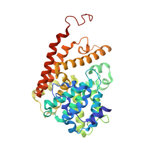
    To better understand the structural origins of inhibitor selectivity of human phosphodieasterase families (PDEs 1-11), here we report the X-ray crystal structure of PDE2 in complex with a highly selective, nanomolar inhibitor (BAY60-7550) at 1.9 Å resolution, and the structure of apo PDE2 at 2.0 Å resolution. The crystal structures reveal that the inhibitor binds to the PDE2 active site by using not only the conserved glutamine-switch mechanism for substrate binding, but also a binding-induced, hydrophobic pocket that was not reported previously. In silico affinity profiling by molecular docking indicates that the inhibitor binding to this pocket contributes significantly to the binding affinity and thereby improves the inhibitor selectivity for PDE2. Our results highlight a structure-based design strategy that exploits the potential binding-induced pockets to achieve higher selectivity in the PDE inhibitor development.


  • Organizational Affiliation

    State Key Laboratory of Genetic Engineering, School of Life Sciences, Fudan University, Shanghai 200433, China.


Macromolecules
Find similar proteins by:  (by identity cutoff)  |  3D Structure
Entity ID: 1
MoleculeChains Sequence LengthOrganismDetailsImage
cGMP-dependent 3',5'-cyclic phosphodiesterase
A, B, C, D
342Homo sapiensMutation(s): 0 
Gene Names: PDE2A
EC: 3.1.4.17
UniProt & NIH Common Fund Data Resources
Find proteins for O00408 (Homo sapiens)
Explore O00408 
Go to UniProtKB:  O00408
PHAROS:  O00408
GTEx:  ENSG00000186642 
Entity Groups  
Sequence Clusters30% Identity50% Identity70% Identity90% Identity95% Identity100% Identity
UniProt GroupO00408
Sequence Annotations
Expand
  • Reference Sequence
Small Molecules
Ligands 3 Unique
IDChains Name / Formula / InChI Key2D Diagram3D Interactions
19F
Query on 19F

Download Ideal Coordinates CCD File 
G [auth A],
J [auth B],
M [auth C],
P [auth D]
2-(3,4-dimethoxybenzyl)-7-[(2R,3R)-2-hydroxy-6-phenylhexan-3-yl]-5-methylimidazo[5,1-f][1,2,4]triazin-4(3H)-one
C27 H32 N4 O4
MYTWFJKBZGMYCS-NQIIRXRSSA-N
ZN
Query on ZN

Download Ideal Coordinates CCD File 
E [auth A],
H [auth B],
K [auth C],
N [auth D]
ZINC ION
Zn
PTFCDOFLOPIGGS-UHFFFAOYSA-N
MG
Query on MG

Download Ideal Coordinates CCD File 
F [auth A],
I [auth B],
L [auth C],
O [auth D]
MAGNESIUM ION
Mg
JLVVSXFLKOJNIY-UHFFFAOYSA-N
Binding Affinity Annotations 
IDSourceBinding Affinity
19F Binding MOAD:  4HTX IC50: 4.7 (nM) from 1 assay(s)
PDBBind:  4HTX IC50: 4.7 (nM) from 1 assay(s)
Experimental Data & Validation

Experimental Data

  • Method: X-RAY DIFFRACTION
  • Resolution: 1.90 Å
  • R-Value Free: 0.227 
  • R-Value Work: 0.173 
  • R-Value Observed: 0.175 
  • Space Group: P 1
Unit Cell:
Length ( Å )Angle ( ˚ )
a = 56.26α = 109.59
b = 73.82β = 91.93
c = 92.47γ = 90.88
Software Package:
Software NamePurpose
ADSCdata collection
MOLREPphasing
PHENIXrefinement
iMOSFLMdata reduction

Structure Validation

View Full Validation Report


Ligand Structure Quality Assessment 


Entry History 

Deposition Data

Revision History  (Full details and data files)

  • Version 1.0: 2013-08-28
    Type: Initial release
  • Version 1.1: 2014-12-17
    Changes: Data collection
  • Version 1.2: 2017-11-15
    Changes: Refinement description
  • Version 1.3: 2023-09-20
    Changes: Data collection, Database references, Derived calculations, Refinement description