4HON

Crystal structure of human JMJD2D/KDM4D in complex with an H3K9me3 peptide and 2-oxoglutarate


Experimental Data Snapshot

  • Method: X-RAY DIFFRACTION
  • Resolution: 1.80 Å
  • R-Value Free: 0.214 
  • R-Value Work: 0.189 
  • R-Value Observed: 0.191 

wwPDB Validation   3D Report Full Report


This is version 1.4 of the entry. See complete history


Literature

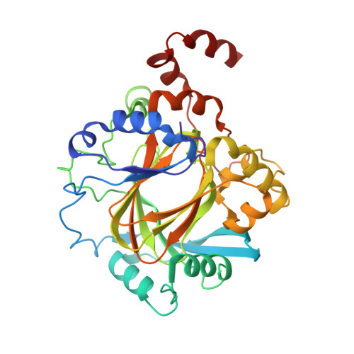
Structural and Functional Analysis of JMJD2D Reveals Molecular Basis for Site-Specific Demethylation among JMJD2 Demethylases.

Krishnan, S.Trievel, R.C.

(2013) Structure 21: 98-108

  • DOI: https://doi.org/10.1016/j.str.2012.10.018
  • Primary Citation of Related Structures:  
    4HON, 4HOO

  • PubMed Abstract: 

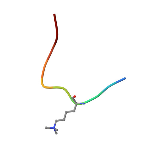
    JMJD2 lysine demethylases (KDMs) participate in diverse genomic processes. Most JMJD2 homologs display dual selectivity toward H3K9me3 and H3K36me3, with the exception of JMJD2D, which is specific for H3K9me3. Here, we report the crystal structures of the JMJD2D⋅2-oxoglutarate⋅H3K9me3 ternary complex and JMJD2D apoenzyme. Utilizing structural alignments with JMJD2A, molecular docking, and kinetic analysis with an array of histone peptide substrates, we elucidate the specific signatures that permit efficient recognition of H3K9me3 by JMJD2A and JMJD2D, and the residues in JMJD2D that occlude H3K36me3 demethylation. Surprisingly, these results reveal that JMJD2A and JMJD2D exhibit subtle yet important differences in H3K9me3 recognition, despite the overall similarity in the substrate-binding conformation. Further, we show that H3T11 phosphorylation abrogates demethylation by JMJD2 KDMs. Together, these studies reveal the molecular basis for JMJD2 site specificity and provide a framework for structure-based design of selective inhibitors of JMJD2 KDMs implicated in disease.


  • Organizational Affiliation

    Department of Biological Chemistry, 1150 West Medical Center Drive, 5301 Medical Science Research Building III, University of Michigan, Ann Arbor, MI 48109, USA.


Macromolecules
Find similar proteins by:  (by identity cutoff)  |  3D Structure
Entity ID: 1
MoleculeChains Sequence LengthOrganismDetailsImage
Lysine-specific demethylase 4D
A, B
330Homo sapiensMutation(s): 0 
Gene Names: JHDM3DJMJD2DKDM4D
EC: 1.14.11
UniProt & NIH Common Fund Data Resources
Find proteins for Q6B0I6 (Homo sapiens)
Explore Q6B0I6 
Go to UniProtKB:  Q6B0I6
PHAROS:  Q6B0I6
GTEx:  ENSG00000186280 
Entity Groups  
Sequence Clusters30% Identity50% Identity70% Identity90% Identity95% Identity100% Identity
UniProt GroupQ6B0I6
Sequence Annotations
Expand
  • Reference Sequence

Find similar proteins by:  Sequence   |   3D Structure  

Entity ID: 2
MoleculeChains Sequence LengthOrganismDetailsImage
Histone H3 PeptideC [auth F],
D [auth G]
10Homo sapiensMutation(s): 0 
UniProt & NIH Common Fund Data Resources
Find proteins for P68431 (Homo sapiens)
Explore P68431 
Go to UniProtKB:  P68431
PHAROS:  P68431
Entity Groups  
Sequence Clusters30% Identity50% Identity70% Identity90% Identity95% Identity100% Identity
UniProt GroupP68431
Sequence Annotations
Expand
  • Reference Sequence
Small Molecules
Ligands 6 Unique
IDChains Name / Formula / InChI Key2D Diagram3D Interactions
AKG
Query on AKG

Download Ideal Coordinates CCD File 
E [auth A],
W [auth B]
2-OXOGLUTARIC ACID
C5 H6 O5
KPGXRSRHYNQIFN-UHFFFAOYSA-N
PDO
Query on PDO

Download Ideal Coordinates CCD File 
GA [auth B],
S [auth A],
T [auth A],
U [auth A],
V [auth A]
1,3-PROPANDIOL
C3 H8 O2
YPFDHNVEDLHUCE-UHFFFAOYSA-N
ZN
Query on ZN

Download Ideal Coordinates CCD File 
G [auth A],
Y [auth B]
ZINC ION
Zn
PTFCDOFLOPIGGS-UHFFFAOYSA-N
NO3
Query on NO3

Download Ideal Coordinates CCD File 
CA [auth B]
DA [auth B]
EA [auth B]
FA [auth B]
K [auth A]
CA [auth B],
DA [auth B],
EA [auth B],
FA [auth B],
K [auth A],
L [auth A],
M [auth A],
N [auth A],
O [auth A],
P [auth A],
Q [auth A],
R [auth A]
NITRATE ION
N O3
NHNBFGGVMKEFGY-UHFFFAOYSA-N
NI
Query on NI

Download Ideal Coordinates CCD File 
F [auth A],
X [auth B]
NICKEL (II) ION
Ni
VEQPNABPJHWNSG-UHFFFAOYSA-N
SCN
Query on SCN

Download Ideal Coordinates CCD File 
AA [auth B]
BA [auth B]
H [auth A]
I [auth A]
J [auth A]
AA [auth B],
BA [auth B],
H [auth A],
I [auth A],
J [auth A],
Z [auth B]
THIOCYANATE ION
C N S
ZMZDMBWJUHKJPS-UHFFFAOYSA-M
Modified Residues  1 Unique
IDChains TypeFormula2D DiagramParent
M3L
Query on M3L
C [auth F],
D [auth G]
L-PEPTIDE LINKINGC9 H21 N2 O2LYS
Experimental Data & Validation

Experimental Data

  • Method: X-RAY DIFFRACTION
  • Resolution: 1.80 Å
  • R-Value Free: 0.214 
  • R-Value Work: 0.189 
  • R-Value Observed: 0.191 
  • Space Group: P 21 21 21
Unit Cell:
Length ( Å )Angle ( ˚ )
a = 72.202α = 90
b = 79.675β = 90
c = 175.938γ = 90
Software Package:
Software NamePurpose
DENZOdata reduction
SCALEPACKdata scaling
REFMACrefinement
PDB_EXTRACTdata extraction
HKL-2000data collection
HKL-2000data reduction
CCP4phasing

Structure Validation

View Full Validation Report


Entry History 

Deposition Data

Revision History  (Full details and data files)

  • Version 1.0: 2012-11-21
    Type: Initial release
  • Version 1.1: 2013-01-02
    Changes: Database references
  • Version 1.2: 2013-01-30
    Changes: Database references
  • Version 1.3: 2017-11-15
    Changes: Refinement description
  • Version 1.4: 2023-09-20
    Changes: Data collection, Database references, Derived calculations, Refinement description