4HMN

Crystal structure of human 17beta-hydroxysteroid dehydrogenase type 5 in complex with (4-(4-Chlorophenyl)piperazin-1-yl)(morpholino)methanone (24)


Experimental Data Snapshot

  • Method: X-RAY DIFFRACTION
  • Resolution: 2.40 Å
  • R-Value Free: 0.297 
  • R-Value Work: 0.219 
  • R-Value Observed: 0.222 

wwPDB Validation   3D Report Full Report


Ligand Structure Quality Assessment 


This is version 1.2 of the entry. See complete history


Literature

Morpholylureas are a new class of potent and selective inhibitors of the type 5 17-beta-hydroxysteroid dehydrogenase (AKR1C3).

Flanagan, J.U.Atwell, G.J.Heinrich, D.M.Brooke, D.G.Silva, S.Rigoreau, L.J.Trivier, E.Turnbull, A.P.Raynham, T.Jamieson, S.M.Denny, W.A.

(2014) Bioorg Med Chem 22: 967-977

  • DOI: https://doi.org/10.1016/j.bmc.2013.12.050
  • Primary Citation of Related Structures:  
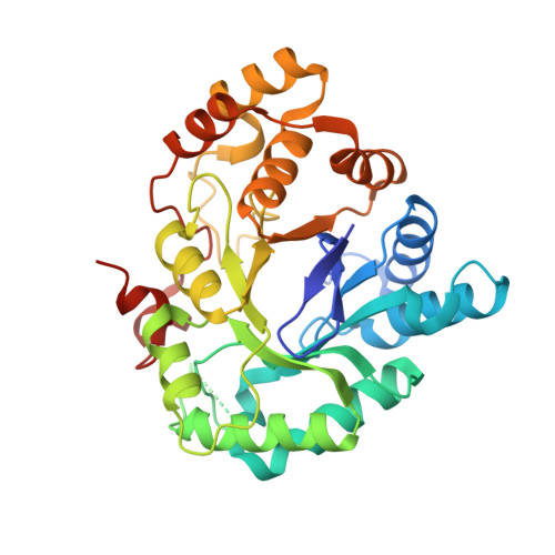
    4HMN

  • PubMed Abstract: 

    Inhibitors of the aldo-keto reductase enzyme AKR1C3 are of interest as potential drugs for leukemia and hormone-related cancers. A series of non-carboxylate morpholino(phenylpiperazin-1-yl)methanones were prepared by palladium-catalysed coupling of substituted phenyl or pyridyl bromides with the known morpholino(piperazin-1-yl)methanone, and shown to be potent (IC50∼100nM) and very isoform-selective inhibitors of AKR1C3. Lipophilic electron-withdrawing substituents on the phenyl ring were positive for activity, as was an H-bond acceptor on the other terminal ring, and the ketone moiety (as a urea) was essential. These structure-activity relationships are consistent with an X-ray structure of a representative compound bound in the AKR1C3 active site, which showed H-bonding between the carbonyl oxygen of the drug and Tyr55 and His117 in the 'oxyanion hole' of the enzyme, with the piperazine bridging unit providing the correct twist to allow the terminal benzene ring to occupy the lipophilic pocket and align with Phe311.


  • Organizational Affiliation

    Auckland Cancer Society Research Centre, The University of Auckland, Private Bag 92019, Auckland 1142, New Zealand; Maurice Wilkins Centre for Molecular Biodiscovery, The University of Auckland, Private Bag 92019, Auckland 1142, New Zealand.


Macromolecules
Find similar proteins by:  (by identity cutoff)  |  3D Structure
Entity ID: 1
MoleculeChains Sequence LengthOrganismDetailsImage
Aldo-keto reductase family 1 member C3331Homo sapiensMutation(s): 0 
Gene Names: AKR1C3DDH1HSD17B5KIAA0119PGFS
EC: 1 (PDB Primary Data), 1.1.1.213 (PDB Primary Data), 1.1.1.112 (PDB Primary Data), 1.1.1.188 (PDB Primary Data), 1.1.1.63 (PDB Primary Data), 1.1.1.64 (PDB Primary Data), 1.3.1.20 (PDB Primary Data)
UniProt & NIH Common Fund Data Resources
Find proteins for P42330 (Homo sapiens)
Explore P42330 
Go to UniProtKB:  P42330
PHAROS:  P42330
GTEx:  ENSG00000196139 
Entity Groups  
Sequence Clusters30% Identity50% Identity70% Identity90% Identity95% Identity100% Identity
UniProt GroupP42330
Sequence Annotations
Expand
  • Reference Sequence
Small Molecules
Ligands 3 Unique
IDChains Name / Formula / InChI Key2D Diagram3D Interactions
NAP
Query on NAP

Download Ideal Coordinates CCD File 
B [auth A]NADP NICOTINAMIDE-ADENINE-DINUCLEOTIDE PHOSPHATE
C21 H28 N7 O17 P3
XJLXINKUBYWONI-NNYOXOHSSA-N
16J
Query on 16J

Download Ideal Coordinates CCD File 
C [auth A][4-(4-chlorophenyl)piperazin-1-yl](morpholin-4-yl)methanone
C15 H20 Cl N3 O2
KDSIRFZJOTZBPT-UHFFFAOYSA-N
EDO
Query on EDO

Download Ideal Coordinates CCD File 
D [auth A]1,2-ETHANEDIOL
C2 H6 O2
LYCAIKOWRPUZTN-UHFFFAOYSA-N
Binding Affinity Annotations 
IDSourceBinding Affinity
16J BindingDB:  4HMN IC50: min: 18, max: 3.00e+4 (nM) from 4 assay(s)
Binding MOAD:  4HMN IC50: 110 (nM) from 1 assay(s)
Experimental Data & Validation

Experimental Data

  • Method: X-RAY DIFFRACTION
  • Resolution: 2.40 Å
  • R-Value Free: 0.297 
  • R-Value Work: 0.219 
  • R-Value Observed: 0.222 
  • Space Group: P 21 21 21
Unit Cell:
Length ( Å )Angle ( ˚ )
a = 57.956α = 90
b = 64.068β = 90
c = 96.404γ = 90
Software Package:
Software NamePurpose
StructureStudiodata collection
PHASERphasing
REFMACrefinement

Structure Validation

View Full Validation Report


Ligand Structure Quality Assessment 


Entry History 

Revision History  (Full details and data files)

  • Version 1.0: 2013-11-13
    Type: Initial release
  • Version 1.1: 2014-02-26
    Changes: Database references
  • Version 1.2: 2023-09-20
    Changes: Data collection, Database references, Derived calculations, Refinement description