Structural basis of Bacillus anthracis MoxXT disruption and the modulation of MoxT ribonuclease activity by rationally designed peptides.
Verma, S., Kumar, S., Gupta, V.P., Gourinath, S., Bhatnagar, S., Bhatnagar, R.(2015) J Biomol Struct Dyn 33: 606-624
- PubMed: 24650157
- DOI: https://doi.org/10.1080/07391102.2014.899924
- Primary Citation of Related Structures:
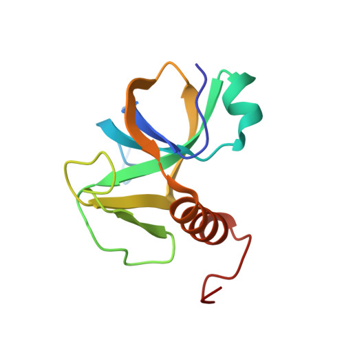
4HKE - PubMed Abstract:
Bacillus anthracis MoxXT is a Type II proteic Toxin-Antitoxin (TA) module wherein MoxT is a ribonuclease that cleaves RNA specifically while MoxX interacts with MoxT and inhibits its activity. Disruption of the TA interaction has been proposed as a novel antibacterial strategy. Peptides, either based on antitoxin sequence or rationally designed, have previously been reported to disrupt the MoxXT interaction but cause a decrease in MoxT ribonuclease activity. In the present study, we report the crystal structure of MoxT, and the effect of several peptides in disrupting the MoxXT interaction as well as augmentation of MoxT ribonuclease activity by binding to MoxT in vitro. Docking studies on the peptides were carried out in order to explain the observed structure activity relationships. The peptides with ribonuclease augmentation activity possess a distinct structure and are proposed to bind to a distinct site on MoxT. The docking of the active peptides with MoxT showed that they possess an aromatic group that occupies a conserved hydrophobic pocket. Additionally, the peptides inducing high ribonuclease activity were anchored by a negatively charged group near a cluster of positively charged residues present near the pocket. Our study provides a structural basis and rationale for the observed properties of the peptides and may aid the development of small molecules to disrupt the TA interaction.
Organizational Affiliation:
a Laboratory of Genetic Engineering and Molecular Biology, School of Biotechnology , Jawaharlal Nehru University , Room No. 102, New Delhi 110067 , India.