4HG3

Structural insights into yeast Nit2: wild-type yeast Nit2 in complex with alpha-ketoglutarate


Experimental Data Snapshot

  • Method: X-RAY DIFFRACTION
  • Resolution: 1.93 Å
  • R-Value Free: 0.227 
  • R-Value Work: 0.197 
  • R-Value Observed: 0.198 

wwPDB Validation   3D Report Full Report


This is version 1.3 of the entry. See complete history


Literature

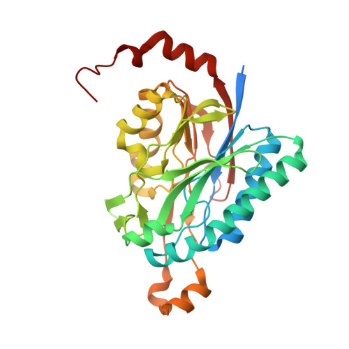
Structures of enzyme-intermediate complexes of yeast Nit2: insights into its catalytic mechanism and different substrate specificity compared with mammalian Nit2

Liu, H.Gao, Y.Zhang, M.Qiu, X.Cooper, A.J.L.Niu, L.Teng, M.

(2013) Acta Crystallogr D Biol Crystallogr 69: 1470-1481

  • DOI: https://doi.org/10.1107/S0907444913009347
  • Primary Citation of Related Structures:  
    4H5U, 4HG3, 4HG5, 4HGD

  • PubMed Abstract: 

    The Nit (nitrilase-like) protein subfamily constitutes branch 10 of the nitrilase superfamily. Nit proteins are widely distributed in nature. Mammals possess two members of the Nit subfamily, namely Nit1 and Nit2. Based on sequence similarity, yeast Nit2 (yNit2) is a homologue of mouse Nit1, a tumour-suppressor protein whose substrate specificity is not yet known. Previous studies have shown that mammalian Nit2 (also a putative tumour suppressor) is identical to ω-amidase, an enzyme that catalyzes the hydrolysis of α-ketoglutaramate (α-KGM) and α-ketosuccinamate (α-KSM) to α-ketoglutarate (α-KG) and oxaloacetate (OA), respectively. In the present study, crystal structures of wild-type (WT) yNit2 and of WT yNit2 in complex with α-KG and with OA were determined. In addition, the crystal structure of the C169S mutant of yNit2 (yNit2-C169S) in complex with an endogenous molecule of unknown structure was also solved. Analysis of the structures revealed that α-KG and OA are covalently bound to Cys169 by the formation of a thioester bond between the sulfhydryl group of the cysteine residue and the γ-carboxyl group of α-KG or the β-carboxyl group of OA, reflecting the presumed reaction intermediates. However, an enzymatic assay suggests that α-KGM is a relatively poor substrate of yNit2. Finally, a ligand was found in the active site of yNit2-C169S that may be a natural substrate of yNit2 or an endogenous regulator of enzyme activity. These crystallographic analyses provide information on the mode of substrate/ligand binding at the active site of yNit2 and insights into the catalytic mechanism. These findings suggest that yNit2 may have broad biological roles in yeast, especially in regard to nitrogen homeostasis, and provide a framework for the elucidation of the substrate specificity and biological role of mammalian Nit1.


  • Organizational Affiliation

    Hefei National Laboratory for Physical Sciences at the Microscale and School of Life Science, University of Science and Technology of China, Hefei, Anhui 230026, People's Republic of China.


Macromolecules
Find similar proteins by:  (by identity cutoff)  |  3D Structure
Entity ID: 1
MoleculeChains Sequence LengthOrganismDetailsImage
Probable hydrolase NIT2
A, B, C, D
341Saccharomyces cerevisiae S288CMutation(s): 0 
Gene Names: NIT2YJL126WJ0706
EC: 3.5
UniProt
Find proteins for P47016 (Saccharomyces cerevisiae (strain ATCC 204508 / S288c))
Explore P47016 
Go to UniProtKB:  P47016
Entity Groups  
Sequence Clusters30% Identity50% Identity70% Identity90% Identity95% Identity100% Identity
UniProt GroupP47016
Sequence Annotations
Expand
  • Reference Sequence
Small Molecules
Ligands 3 Unique
IDChains Name / Formula / InChI Key2D Diagram3D Interactions
AKG
Query on AKG

Download Ideal Coordinates CCD File 
BA [auth D],
E [auth A],
O [auth B],
W [auth C]
2-OXOGLUTARIC ACID
C5 H6 O5
KPGXRSRHYNQIFN-UHFFFAOYSA-N
CAC
Query on CAC

Download Ideal Coordinates CCD File 
AA [auth C]
EA [auth D]
M [auth A]
N [auth A]
U [auth B]
AA [auth C],
EA [auth D],
M [auth A],
N [auth A],
U [auth B],
V [auth B],
Z [auth C]
CACODYLATE ION
C2 H6 As O2
OGGXGZAMXPVRFZ-UHFFFAOYSA-M
GOL
Query on GOL

Download Ideal Coordinates CCD File 
CA [auth D]
DA [auth D]
F [auth A]
G [auth A]
H [auth A]
CA [auth D],
DA [auth D],
F [auth A],
G [auth A],
H [auth A],
I [auth A],
J [auth A],
K [auth A],
L [auth A],
P [auth B],
Q [auth B],
R [auth B],
S [auth B],
T [auth B],
X [auth C],
Y [auth C]
GLYCEROL
C3 H8 O3
PEDCQBHIVMGVHV-UHFFFAOYSA-N
Experimental Data & Validation

Experimental Data

  • Method: X-RAY DIFFRACTION
  • Resolution: 1.93 Å
  • R-Value Free: 0.227 
  • R-Value Work: 0.197 
  • R-Value Observed: 0.198 
  • Space Group: P 1 21 1
Unit Cell:
Length ( Å )Angle ( ˚ )
a = 65.211α = 90
b = 125.653β = 95.3
c = 77.964γ = 90
Software Package:
Software NamePurpose
Blu-Icedata collection
MOLREPphasing
REFMACrefinement
HKL-2000data reduction
HKL-2000data scaling

Structure Validation

View Full Validation Report


Entry History 

Deposition Data

Revision History  (Full details and data files)

  • Version 1.0: 2013-07-31
    Type: Initial release
  • Version 1.1: 2013-08-07
    Changes: Structure summary
  • Version 1.2: 2014-01-22
    Changes: Database references
  • Version 1.3: 2023-11-08
    Changes: Data collection, Database references, Derived calculations, Refinement description