4H75

Crystal structure of human Spindlin1 in complex with a histone H3K4(me3) peptide


Experimental Data Snapshot

  • Method: X-RAY DIFFRACTION
  • Resolution: 2.10 Å
  • R-Value Free: 0.218 
  • R-Value Work: 0.181 
  • R-Value Observed: 0.183 

wwPDB Validation   3D Report Full Report


This is version 1.3 of the entry. See complete history


Literature

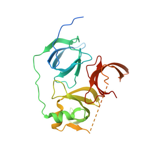
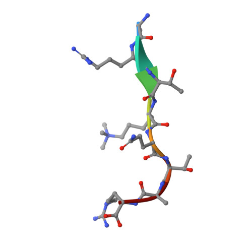
Distinct mode of methylated lysine-4 of histone H3 recognition by tandem tudor-like domains of Spindlin1.

Yang, N.Wang, W.Wang, Y.Wang, M.Zhao, Q.Rao, Z.Zhu, B.Xu, R.M.

(2012) Proc Natl Acad Sci U S A 109: 17954-17959

  • DOI: https://doi.org/10.1073/pnas.1208517109
  • Primary Citation of Related Structures:  
    4H75

  • PubMed Abstract: 

    Recognition of methylated histone tail lysine residues by tudor domains plays important roles in epigenetic control of gene expression and DNA damage response. Previous studies revealed the binding of methyllysine in a cage of aromatic residues, but the molecular mechanism by which the sequence specificity for surrounding histone tail residues is achieved remains poorly understood. In the crystal structure of a trimethylated histone H3 lysine 4 (H3K4) peptide bound to the tudor-like domains of Spindlin1 presented here, an atypical mode of methyllysine recognition by an aromatic pocket of Spindlin1 is observed. Furthermore, the histone sequence is recognized in a distinct manner involving the amino terminus and a pair of arginine residues of histone H3, and disruption of the binding impaired stimulation of pre-RNA expression by Spindlin1. Our analysis demonstrates considerable diversities of methyllysine recognition and sequence-specific binding of histone tails by tudor domains, and the revelation furthers the understanding of tudor domain proteins in deciphering epigenetic marks on histone tails.


  • Organizational Affiliation

    National Laboratory of Biomacromolecules, Institute of Biophysics, Chinese Academy of Sciences, Beijing 100101, China.


Macromolecules
Find similar proteins by:  (by identity cutoff)  |  3D Structure
Entity ID: 1
MoleculeChains Sequence LengthOrganismDetailsImage
Spindlin-1238Homo sapiensMutation(s): 0 
Gene Names: SPIN1OCRSPIN
UniProt & NIH Common Fund Data Resources
Find proteins for Q9Y657 (Homo sapiens)
Explore Q9Y657 
Go to UniProtKB:  Q9Y657
PHAROS:  Q9Y657
GTEx:  ENSG00000106723 
Entity Groups  
Sequence Clusters30% Identity50% Identity70% Identity90% Identity95% Identity100% Identity
UniProt GroupQ9Y657
Sequence Annotations
Expand
  • Reference Sequence

Find similar proteins by:  Sequence   |   3D Structure  

Entity ID: 2
MoleculeChains Sequence LengthOrganismDetailsImage
Histone H38Homo sapiensMutation(s): 0 
UniProt
Find proteins for Q5TEC6 (Homo sapiens)
Explore Q5TEC6 
Go to UniProtKB:  Q5TEC6
Entity Groups  
UniProt GroupQ5TEC6
Sequence Annotations
Expand
  • Reference Sequence
Experimental Data & Validation

Experimental Data

  • Method: X-RAY DIFFRACTION
  • Resolution: 2.10 Å
  • R-Value Free: 0.218 
  • R-Value Work: 0.181 
  • R-Value Observed: 0.183 
  • Space Group: P 21 21 2
Unit Cell:
Length ( Å )Angle ( ˚ )
a = 122.429α = 90
b = 42.685β = 90
c = 50.307γ = 90
Software Package:
Software NamePurpose
ADSCdata collection
MOLREPphasing
PHENIXrefinement
HKL-2000data reduction
HKL-2000data scaling

Structure Validation

View Full Validation Report


Entry History 

Deposition Data

Revision History  (Full details and data files)

  • Version 1.0: 2012-10-03
    Type: Initial release
  • Version 1.1: 2012-10-31
    Changes: Database references
  • Version 1.2: 2012-11-14
    Changes: Database references
  • Version 1.3: 2023-09-20
    Changes: Data collection, Database references, Derived calculations, Refinement description