4H4A

Crystal structure of the C-terminal domain of Drosophila melanogaster Zucchini


Experimental Data Snapshot

  • Method: X-RAY DIFFRACTION
  • Resolution: 2.20 Å
  • R-Value Free: 0.255 
  • R-Value Work: 0.218 
  • R-Value Observed: 0.222 

wwPDB Validation   3D Report Full Report


This is version 1.2 of the entry. See complete history


Literature

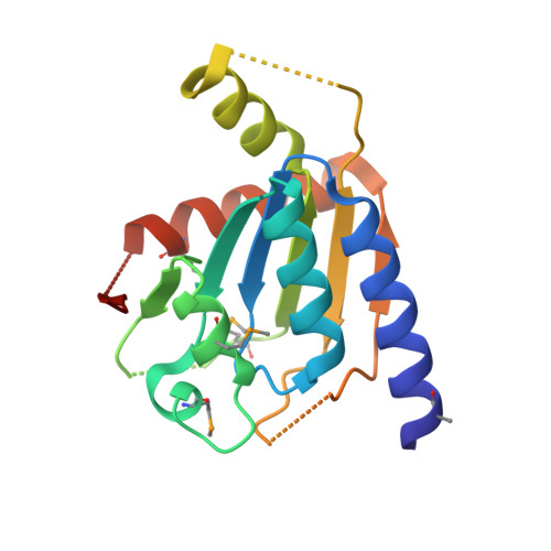
Crystal structure of the primary piRNA biogenesis factor Zucchini reveals similarity to the bacterial PLD endonuclease Nuc.

Voigt, F.Reuter, M.Kasaruho, A.Schulz, E.C.Pillai, R.S.Barabas, O.

(2012) RNA 18: 2128-2134

  • DOI: https://doi.org/10.1261/rna.034967.112
  • Primary Citation of Related Structures:  
    4H4A

  • PubMed Abstract: 

    Piwi-interacting RNAs (piRNAs) are a gonad-specific class of small RNAs that associate with the Piwi clade of Argonaute proteins and play a key role in transposon silencing in animals. Since biogenesis of piRNAs is independent of the double-stranded RNA-processing enzyme Dicer, an alternative nuclease that can process single-stranded RNA transcripts has been long sought. A Phospholipase D-like protein, Zucchini, that is essential for piRNA processing has been proposed to be a nuclease acting in piRNA biogenesis. Here we describe the crystal structure of Zucchini from Drosophila melanogaster and show that it is very similar to the bacterial endonuclease, Nuc. The structure also reveals that homodimerization induces major conformational changes assembling the active site. The active site is situated on the dimer interface at the bottom of a narrow groove that can likely accommodate single-stranded nucleic acid substrates. Furthermore, biophysical analysis identifies protein segments essential for dimerization and provides insights into regulation of Zucchini's activity.


Macromolecules
Find similar proteins by:  (by identity cutoff)  |  3D Structure
Entity ID: 1
MoleculeChains Sequence LengthOrganismDetailsImage
Mitochondrial cardiolipin hydrolase169Drosophila melanogasterMutation(s): 0 
Gene Names: zucCG12314
EC: 3.1.4
UniProt
Find proteins for Q9VKD7 (Drosophila melanogaster)
Explore Q9VKD7 
Go to UniProtKB:  Q9VKD7
Entity Groups  
Sequence Clusters30% Identity50% Identity70% Identity90% Identity95% Identity100% Identity
UniProt GroupQ9VKD7
Sequence Annotations
Expand
  • Reference Sequence
Small Molecules
Modified Residues  1 Unique
IDChains TypeFormula2D DiagramParent
MSE
Query on MSE
A
L-PEPTIDE LINKINGC5 H11 N O2 SeMET
Experimental Data & Validation

Experimental Data

  • Method: X-RAY DIFFRACTION
  • Resolution: 2.20 Å
  • R-Value Free: 0.255 
  • R-Value Work: 0.218 
  • R-Value Observed: 0.222 
  • Space Group: P 1 21 1
Unit Cell:
Length ( Å )Angle ( ˚ )
a = 35.64α = 90
b = 51.28β = 107.63
c = 40.97γ = 90
Software Package:
Software NamePurpose
Auto-Rickshawphasing
SHELXmodel building
PHENIXrefinement
XDSdata reduction
XSCALEdata scaling
SHELXphasing

Structure Validation

View Full Validation Report


Entry History 

Deposition Data

Revision History  (Full details and data files)

  • Version 1.0: 2012-11-07
    Type: Initial release
  • Version 1.1: 2012-12-19
    Changes: Database references
  • Version 1.2: 2017-11-15
    Changes: Refinement description