4H1W

E1 structure of the (SR) Ca2+-ATPase in complex with Sarcolipin


Experimental Data Snapshot

  • Method: X-RAY DIFFRACTION
  • Resolution: 3.10 Å
  • R-Value Free: 0.247 
  • R-Value Work: 0.191 
  • R-Value Observed: 0.194 

wwPDB Validation   3D Report Full Report


Ligand Structure Quality Assessment 


This is version 1.3 of the entry. See complete history


Literature

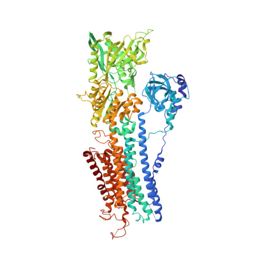
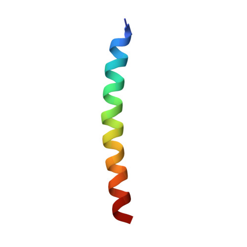
The sarcolipin-bound calcium pump stabilizes calcium sites exposed to the cytoplasm.

Winther, A.M.Bublitz, M.Karlsen, J.L.Moller, J.V.Hansen, J.B.Nissen, P.Buch-Pedersen, M.J.

(2013) Nature 495: 265-269

  • DOI: https://doi.org/10.1038/nature11900
  • Primary Citation of Related Structures:  
    4H1W

  • PubMed Abstract: 

    The contraction and relaxation of muscle cells is controlled by the successive rise and fall of cytosolic Ca(2+), initiated by the release of Ca(2+) from the sarcoplasmic reticulum and terminated by re-sequestration of Ca(2+) into the sarcoplasmic reticulum as the main mechanism of Ca(2+) removal. Re-sequestration requires active transport and is catalysed by the sarcoplasmic reticulum Ca(2+)-ATPase (SERCA), which has a key role in defining the contractile properties of skeletal and heart muscle tissue. The activity of SERCA is regulated by two small, homologous membrane proteins called phospholamban (PLB, also known as PLN) and sarcolipin (SLN). Detailed structural information explaining this regulatory mechanism has been lacking, and the structural features defining the pathway through which cytoplasmic Ca(2+) enters the intramembranous binding sites of SERCA have remained unknown. Here we report the crystal structure of rabbit SERCA1a (also known as ATP2A1) in complex with SLN at 3.1 Å resolution. The regulatory SLN traps the Ca(2+)-ATPase in a previously undescribed E1 state, with exposure of the Ca(2+) sites through an open cytoplasmic pathway stabilized by Mg(2+). The structure suggests a mechanism for selective Ca(2+) loading and activation of SERCA, and provides new insight into how SLN and PLB inhibition arises from stabilization of this E1 intermediate state without bound Ca(2+). These findings may prove useful in studying how autoinhibitory domains of other ion pumps modulate transport across biological membranes.


  • Organizational Affiliation

    Pcovery, Thorvaldsensvej 57, DK-1871 Frederiksberg, Denmark.


Macromolecules
Find similar proteins by:  (by identity cutoff)  |  3D Structure
Entity ID: 1
MoleculeChains Sequence LengthOrganismDetailsImage
SERCA1a994Oryctolagus cuniculusMutation(s): 0 
EC: 3.6.3.8
Membrane Entity: Yes 
UniProt
Find proteins for P04191 (Oryctolagus cuniculus)
Explore P04191 
Go to UniProtKB:  P04191
Entity Groups  
Sequence Clusters30% Identity50% Identity70% Identity90% Identity95% Identity100% Identity
UniProt GroupP04191
Sequence Annotations
Expand
  • Reference Sequence
Find similar proteins by:  (by identity cutoff)  |  3D Structure
Entity ID: 2
MoleculeChains Sequence LengthOrganismDetailsImage
Sarcolipin31Oryctolagus cuniculusMutation(s): 0 
Membrane Entity: Yes 
UniProt
Find proteins for P42532 (Oryctolagus cuniculus)
Explore P42532 
Go to UniProtKB:  P42532
Entity Groups  
Sequence Clusters30% Identity50% Identity70% Identity90% Identity95% Identity100% Identity
UniProt GroupP42532
Sequence Annotations
Expand
  • Reference Sequence
Experimental Data & Validation

Experimental Data

  • Method: X-RAY DIFFRACTION
  • Resolution: 3.10 Å
  • R-Value Free: 0.247 
  • R-Value Work: 0.191 
  • R-Value Observed: 0.194 
  • Space Group: P 21 2 21
Unit Cell:
Length ( Å )Angle ( ˚ )
a = 85.87α = 90
b = 139.54β = 90
c = 141.56γ = 90
Software Package:
Software NamePurpose
XSCALEdata scaling
PHENIXrefinement
PDB_EXTRACTdata extraction
RemDAqdata collection
XDSdata reduction
PHASERphasing

Structure Validation

View Full Validation Report


Ligand Structure Quality Assessment 


Entry History 

Deposition Data

Revision History  (Full details and data files)

  • Version 1.0: 2013-03-06
    Type: Initial release
  • Version 1.1: 2013-03-27
    Changes: Database references
  • Version 1.2: 2017-11-15
    Changes: Refinement description
  • Version 1.3: 2024-02-28
    Changes: Data collection, Database references, Derived calculations