4H0E

Crystal Structure of mutant ORR3 in complex with NTD of AraR


Experimental Data Snapshot

  • Method: X-RAY DIFFRACTION
  • Resolution: 1.97 Å
  • R-Value Free: 0.253 
  • R-Value Work: 0.219 
  • R-Value Observed: 0.221 

wwPDB Validation   3D Report Full Report


This is version 1.1 of the entry. See complete history


Literature

Spacing between core recognition motifs determines relative orientation of AraR monomers on bipartite operators.

Jain, D.Nair, D.T.

(2013) Nucleic Acids Res 41: 639-647

  • DOI: https://doi.org/10.1093/nar/gks962
  • Primary Citation of Related Structures:  
    4EGY, 4EGZ, 4H0E

  • PubMed Abstract: 

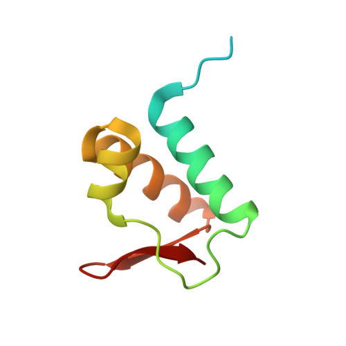
    Transcription factors modulate expression primarily through specific recognition of cognate sequences resident in the promoter region of target genes. AraR (Bacillus subtilis) is a repressor of genes involved in L-arabinose metabolism. It binds to eight different operators present in five different promoters with distinct affinities through a DNA binding domain at the N-terminus. The structures of AraR-NTD in complex with two distinct operators (ORA1 and ORR3) reveal that two monomers bind to one recognition motif (T/ANG) each in the bipartite operators. The structures show that the two recognition motifs are spaced apart by six bases in cases of ORA1 and eight bases in case of ORR3. This increase in the spacing in the operators by two base pairs results in a drastic change in the position and orientation of the second monomer on DNA in the case of ORR3 when compared with ORA1. Because AraR binds to the two operators with distinct affinities to achieve different levels of repression, this observation suggests that the variation in the spacing between core recognition motifs could be a strategy used by this transcription modulator to differentially influence gene expression.


  • Organizational Affiliation

    Laboratory 4, National Centre for Biological Sciences, NCBS-TIFR, UAS-GKVK Campus, Bellary Road, Bangalore, Karnataka 560065, India.


Macromolecules

Find similar proteins by:  (by identity cutoff)  |  3D Structure
Entity ID: 1
MoleculeChains Sequence LengthOrganismDetailsImage
Arabinose metabolism transcriptional repressor
A, B
88Bacillus subtilisMutation(s): 0 
Gene Names: araCaraRBSU33970yvbS
UniProt
Find proteins for P96711 (Bacillus subtilis (strain 168))
Explore P96711 
Go to UniProtKB:  P96711
Entity Groups  
Sequence Clusters30% Identity50% Identity70% Identity90% Identity95% Identity100% Identity
UniProt GroupP96711
Sequence Annotations
Expand
  • Reference Sequence

Find similar nucleic acids by:  Sequence   |   3D Structure  

Entity ID: 2
MoleculeChains LengthOrganismImage
5'-D(*AP*AP*AP*TP*TP*TP*GP*TP*CP*CP*GP*TP*AP*CP*AP*TP*TP*TP*TP*AP*T)-3'C [auth U]21N/A
Sequence Annotations
Expand
  • Reference Sequence

Find similar nucleic acids by:  Sequence   |   3D Structure  

Entity ID: 3
MoleculeChains LengthOrganismImage
5'-D(*TP*AP*TP*AP*AP*AP*AP*TP*GP*TP*AP*CP*GP*GP*AP*CP*AP*AP*AP*TP*T)-3'D [auth T]21N/A
Sequence Annotations
Expand
  • Reference Sequence
Experimental Data & Validation

Experimental Data

  • Method: X-RAY DIFFRACTION
  • Resolution: 1.97 Å
  • R-Value Free: 0.253 
  • R-Value Work: 0.219 
  • R-Value Observed: 0.221 
  • Space Group: C 1 2 1
Unit Cell:
Length ( Å )Angle ( ˚ )
a = 137.8α = 90
b = 42.62β = 114.92
c = 67.44γ = 90
Software Package:
Software NamePurpose
PXIIIdata collection
PHASERphasing
PHENIXrefinement
XDSdata reduction
XDSdata scaling

Structure Validation

View Full Validation Report


Entry History 

Deposition Data

Revision History  (Full details and data files)

  • Version 1.0: 2013-02-06
    Type: Initial release
  • Version 1.1: 2023-11-08
    Changes: Data collection, Database references, Derived calculations, Refinement description