4GZR

Crystal structure of the Mycobacterium tuberculosis H37Rv EsxOP (Rv2346c-Rv2347c) complex in space group C2221


Experimental Data Snapshot

  • Method: X-RAY DIFFRACTION
  • Resolution: 2.55 Å
  • R-Value Free: 0.233 
  • R-Value Work: 0.199 
  • R-Value Observed: 0.202 

wwPDB Validation   3D Report Full Report


This is version 1.3 of the entry. See complete history


Literature

Heterologous expression of mycobacterial Esx complexes in Escherichia coli for structural studies is facilitated by the use of maltose binding protein fusions.

Arbing, M.A.Chan, S.Harris, L.Kuo, E.Zhou, T.T.Ahn, C.J.Nguyen, L.He, Q.Lu, J.Menchavez, P.T.Shin, A.Holton, T.Sawaya, M.R.Cascio, D.Eisenberg, D.

(2013) PLoS One 8: e81753-e81753

  • DOI: https://doi.org/10.1371/journal.pone.0081753
  • Primary Citation of Related Structures:  
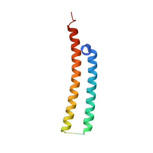
    3OGI, 3Q4H, 4GZR, 4I0X

  • PubMed Abstract: 

    The expression of heteroligomeric protein complexes for structural studies often requires a special coexpression strategy. The reason is that the solubility and proper folding of each subunit of the complex requires physical association with other subunits of the complex. The genomes of pathogenic mycobacteria encode many small protein complexes, implicated in bacterial fitness and pathogenicity, whose characterization may be further complicated by insolubility upon expression in Escherichia coli, the most common heterologous protein expression host. As protein fusions have been shown to dramatically affect the solubility of the proteins to which they are fused, we evaluated the ability of maltose binding protein fusions to produce mycobacterial Esx protein complexes. A single plasmid expression strategy using an N-terminal maltose binding protein fusion to the CFP-10 homolog proved effective in producing soluble Esx protein complexes, as determined by a small-scale expression and affinity purification screen, and coupled with intracellular proteolytic cleavage of the maltose binding protein moiety produced protein complexes of sufficient purity for structural studies. In comparison, the expression of complexes with hexahistidine affinity tags alone on the CFP-10 subunits failed to express in amounts sufficient for biochemical characterization. Using this strategy, six mycobacterial Esx complexes were expressed, purified to homogeneity, and subjected to crystallization screening and the crystal structures of the Mycobacterium abscessus EsxEF, M. smegmatis EsxGH, and M. tuberculosis EsxOP complexes were determined. Maltose binding protein fusions are thus an effective method for production of Esx complexes and this strategy may be applicable for production of other protein complexes.


  • Organizational Affiliation

    UCLA-DOE Institute for Genomics and Proteomics, University of California Los Angeles, Los Angeles, California, United States of America.


Macromolecules
Find similar proteins by:  (by identity cutoff)  |  3D Structure
Entity ID: 1
MoleculeChains Sequence LengthOrganismDetailsImage
ESAT-6-like protein 6
A, C
101Mycobacterium tuberculosis H37RvMutation(s): 0 
Gene Names: MT2411MTCY98.15cRv2346c
UniProt
Find proteins for P9WNI7 (Mycobacterium tuberculosis (strain ATCC 25618 / H37Rv))
Explore P9WNI7 
Go to UniProtKB:  P9WNI7
Entity Groups  
Sequence Clusters30% Identity50% Identity70% Identity90% Identity95% Identity100% Identity
UniProt GroupP9WNI7
Sequence Annotations
Expand
  • Reference Sequence
Find similar proteins by:  (by identity cutoff)  |  3D Structure
Entity ID: 2
MoleculeChains Sequence LengthOrganismDetailsImage
ESAT-6-like protein 7
B, D
99Mycobacterium tuberculosis H37RvMutation(s): 0 
Gene Names: MT2412MTCY98.16cRv2347c
UniProt
Find proteins for P9WNI5 (Mycobacterium tuberculosis (strain ATCC 25618 / H37Rv))
Explore P9WNI5 
Go to UniProtKB:  P9WNI5
Entity Groups  
Sequence Clusters30% Identity50% Identity70% Identity90% Identity95% Identity100% Identity
UniProt GroupP9WNI5
Sequence Annotations
Expand
  • Reference Sequence
Small Molecules
Ligands 1 Unique
IDChains Name / Formula / InChI Key2D Diagram3D Interactions
SO4
Query on SO4

Download Ideal Coordinates CCD File 
E [auth D]SULFATE ION
O4 S
QAOWNCQODCNURD-UHFFFAOYSA-L
Experimental Data & Validation

Experimental Data

  • Method: X-RAY DIFFRACTION
  • Resolution: 2.55 Å
  • R-Value Free: 0.233 
  • R-Value Work: 0.199 
  • R-Value Observed: 0.202 
  • Space Group: C 2 2 21
Unit Cell:
Length ( Å )Angle ( ˚ )
a = 76.1α = 90
b = 112.07β = 90
c = 95.31γ = 90
Software Package:
Software NamePurpose
XSCALEdata scaling
PHASERphasing
PHENIXrefinement
PDB_EXTRACTdata extraction
ADSCdata collection
XDSdata reduction

Structure Validation

View Full Validation Report


Entry History 

Revision History  (Full details and data files)

  • Version 1.0: 2012-09-26
    Type: Initial release
  • Version 1.1: 2012-10-03
    Changes: Other
  • Version 1.2: 2014-05-21
    Changes: Database references
  • Version 1.3: 2023-09-13
    Changes: Data collection, Database references, Derived calculations, Refinement description