4GZN

Mouse ZFP57 zinc fingers in complex with methylated DNA


Experimental Data Snapshot

  • Method: X-RAY DIFFRACTION
  • Resolution: 0.99 Å
  • R-Value Free: 0.140 
  • R-Value Work: 0.131 
  • R-Value Observed: 0.132 

wwPDB Validation   3D Report Full Report


This is version 1.2 of the entry. See complete history


Literature

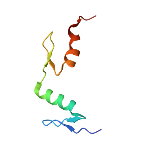
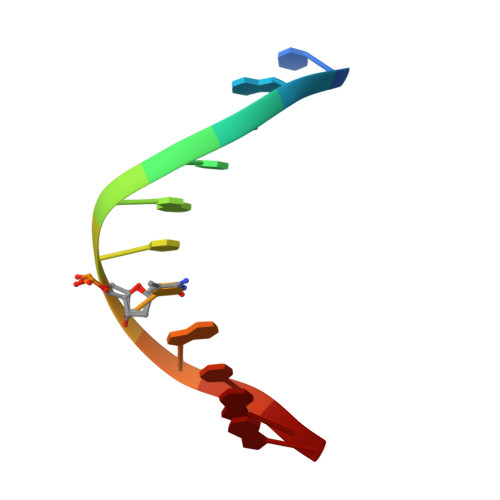
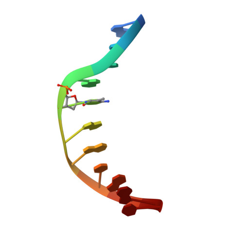
An atomic model of Zfp57 recognition of CpG methylation within a specific DNA sequence.

Liu, Y.Toh, H.Sasaki, H.Zhang, X.Cheng, X.

(2012) Genes Dev 26: 2374-2379

  • DOI: https://doi.org/10.1101/gad.202200.112
  • Primary Citation of Related Structures:  
    4GZN

  • PubMed Abstract: 

    Zinc finger transcription factor Zfp57 recognizes the methylated CpG within the TGCCGC element. We determined the structure of the DNA-binding domain of Zfp57, consisting of two adjacent zinc fingers, in complex with fully methylated DNA at 1.0 Å resolution. The first zinc finger contacts the 5' half (TGC), and the second recognizes the 3' half (CGC) of the recognition sequence. Zfp57 recognizes the two 5-methylcytosines (5mCs) asymmetrically: One involves hydrophobic interactions with Arg178, which also interacts with the neighboring 3' guanine and forms a 5mC-Arg-G interaction, while the other involves a layer of ordered water molecules. Two point mutations in patients with transient neonatal diabetes abolish DNA-binding activity. Zfp57 has reduced binding affinity for unmodified DNA and the oxidative products of 5mC.


  • Organizational Affiliation

    Department of Biochemistry, Emory University School of Medicine, Atlanta, GA 30322, USA.


Macromolecules

Find similar proteins by:  (by identity cutoff)  |  3D Structure
Entity ID: 3
MoleculeChains Sequence LengthOrganismDetailsImage
Zinc finger protein 5760Mus musculusMutation(s): 0 
Gene Names: Zfp57
UniProt
Find proteins for Q8C6P8 (Mus musculus)
Explore Q8C6P8 
Go to UniProtKB:  Q8C6P8
Entity Groups  
Sequence Clusters30% Identity50% Identity70% Identity90% Identity95% Identity100% Identity
UniProt GroupQ8C6P8
Sequence Annotations
Expand
  • Reference Sequence

Find similar nucleic acids by:  Sequence   |   3D Structure  

Entity ID: 1
MoleculeChains LengthOrganismImage
DNA (5'-D(*TP*AP*TP*TP*GP*CP*(5CM)P*GP*CP*AP*G)-3')11N/A
Sequence Annotations
Expand
  • Reference Sequence

Find similar nucleic acids by:  Sequence   |   3D Structure  

Entity ID: 2
MoleculeChains LengthOrganismImage
DNA (5'-D(*AP*CP*TP*GP*(5CM)P*GP*GP*CP*AP*AP*T)-3')11N/A
Sequence Annotations
Expand
  • Reference Sequence
Experimental Data & Validation

Experimental Data

  • Method: X-RAY DIFFRACTION
  • Resolution: 0.99 Å
  • R-Value Free: 0.140 
  • R-Value Work: 0.131 
  • R-Value Observed: 0.132 
  • Space Group: C 2 2 21
Unit Cell:
Length ( Å )Angle ( ˚ )
a = 40.218α = 90
b = 60.641β = 90
c = 96.119γ = 90
Software Package:
Software NamePurpose
HKL-2000data collection
PHENIXmodel building
PHENIXrefinement
HKL-2000data reduction
HKL-2000data scaling
PHENIXphasing

Structure Validation

View Full Validation Report


Entry History 

Deposition Data

Revision History  (Full details and data files)

  • Version 1.0: 2012-10-17
    Type: Initial release
  • Version 1.1: 2012-11-21
    Changes: Database references
  • Version 1.2: 2024-02-28
    Changes: Data collection, Database references, Derived calculations