4GVL

Crystal Structure of the GsuK RCK domain


Experimental Data Snapshot

  • Method: X-RAY DIFFRACTION
  • Resolution: 3.00 Å
  • R-Value Free: 0.262 
  • R-Value Work: 0.228 
  • R-Value Observed: 0.229 

wwPDB Validation   3D Report Full Report


Ligand Structure Quality Assessment 


This is version 1.1 of the entry. See complete history


Literature

Distinct gating mechanisms revealed by the structures of a multi-ligand gated K(+) channel.

Kong, C.Zeng, W.Ye, S.Chen, L.Sauer, D.B.Lam, Y.Derebe, M.G.Jiang, Y.

(2012) Elife 1: e00184-e00184

  • DOI: https://doi.org/10.7554/eLife.00184
  • Primary Citation of Related Structures:  
    4GVL, 4GX0, 4GX1, 4GX2, 4GX5

  • PubMed Abstract: 

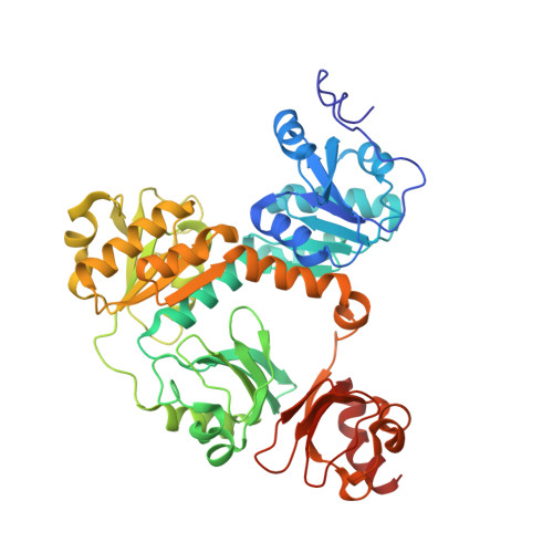
    The gating ring-forming RCK domain regulates channel gating in response to various cellular chemical stimuli in eukaryotic Slo channel families and the majority of ligand-gated prokaryotic K(+) channels and transporters. Here we present structural and functional studies of a dual RCK-containing, multi-ligand gated K(+) channel from Geobacter sulfurreducens, named GsuK. We demonstrate that ADP and NAD(+) activate the GsuK channel, whereas Ca(2+) serves as an allosteric inhibitor. Multiple crystal structures elucidate the structural basis of multi-ligand gating in GsuK, and also reveal a unique ion conduction pore with segmented inner helices. Structural comparison leads us to propose a novel pore opening mechanics that is distinct from other K(+) channels.DOI:http://dx.doi.org/10.7554/eLife.00184.001.


  • Organizational Affiliation

    Department of Physiology , University of Texas Southwestern Medical Center , Dallas , United States.


Macromolecules
Find similar proteins by:  (by identity cutoff)  |  3D Structure
Entity ID: 1
MoleculeChains Sequence LengthOrganismDetailsImage
TrkA domain protein
A, B, C, D
462Geobacter sulfurreducens PCAMutation(s): 0 
Gene Names: GSU0527
Membrane Entity: Yes 
UniProt
Find proteins for Q74FS9 (Geobacter sulfurreducens (strain ATCC 51573 / DSM 12127 / PCA))
Explore Q74FS9 
Go to UniProtKB:  Q74FS9
Entity Groups  
Sequence Clusters30% Identity50% Identity70% Identity90% Identity95% Identity100% Identity
UniProt GroupQ74FS9
Sequence Annotations
Expand
  • Reference Sequence
Small Molecules
Ligands 3 Unique
IDChains Name / Formula / InChI Key2D Diagram3D Interactions
AMP
Query on AMP

Download Ideal Coordinates CCD File 
F [auth A],
J [auth B],
M [auth C],
P [auth D]
ADENOSINE MONOPHOSPHATE
C10 H14 N5 O7 P
UDMBCSSLTHHNCD-KQYNXXCUSA-N
ZN
Query on ZN

Download Ideal Coordinates CCD File 
E [auth A],
I [auth B],
L [auth C],
O [auth D]
ZINC ION
Zn
PTFCDOFLOPIGGS-UHFFFAOYSA-N
CA
Query on CA

Download Ideal Coordinates CCD File 
G [auth A],
H [auth A],
K [auth B],
N [auth C]
CALCIUM ION
Ca
BHPQYMZQTOCNFJ-UHFFFAOYSA-N
Experimental Data & Validation

Experimental Data

  • Method: X-RAY DIFFRACTION
  • Resolution: 3.00 Å
  • R-Value Free: 0.262 
  • R-Value Work: 0.228 
  • R-Value Observed: 0.229 
  • Space Group: I 2 2 2
Unit Cell:
Length ( Å )Angle ( ˚ )
a = 110.648α = 90
b = 161.693β = 90
c = 310.127γ = 90
Software Package:
Software NamePurpose
HKL-2000data collection
PHASERphasing
REFMACrefinement
HKL-2000data reduction
HKL-2000data scaling

Structure Validation

View Full Validation Report


Ligand Structure Quality Assessment 


Entry History 

Deposition Data

Revision History  (Full details and data files)

  • Version 1.0: 2012-12-26
    Type: Initial release
  • Version 1.1: 2023-09-13
    Changes: Data collection, Database references, Derived calculations, Refinement description