4GLX

DNA ligase A in complex with inhibitor


Experimental Data Snapshot

  • Method: X-RAY DIFFRACTION
  • Resolution: 1.90 Å
  • R-Value Free: 0.250 
  • R-Value Work: 0.201 
  • R-Value Observed: 0.203 

wwPDB Validation   3D Report Full Report


Ligand Structure Quality Assessment 


This is version 1.3 of the entry. See complete history


Literature

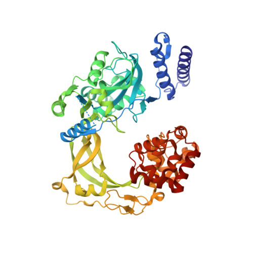
Structure-guided design, synthesis and biological evaluation of novel DNA ligase inhibitors with in vitro and in vivo anti-staphylococcal activity.

Surivet, J.P.Lange, R.Hubschwerlen, C.Keck, W.Specklin, J.L.Ritz, D.Bur, D.Locher, H.Seiler, P.Strasser, D.S.Prade, L.Kohl, C.Schmitt, C.Chapoux, G.Ilhan, E.Ekambaram, N.Athanasiou, A.Knezevic, A.Sabato, D.Chambovey, A.Gaertner, M.Enderlin, M.Boehme, M.Sippel, V.Wyss, P.

(2012) Bioorg Med Chem Lett 22: 6705-6711

  • DOI: https://doi.org/10.1016/j.bmcl.2012.08.094
  • Primary Citation of Related Structures:  
    4GLW, 4GLX

  • PubMed Abstract: 

    A series of 2-amino-[1,8]-naphthyridine-3-carboxamides (ANCs) with potent inhibition of bacterial NAD(+)-dependent DNA ligases (LigAs) evolved from a 2,4-diaminopteridine derivative discovered by HTS. The design was guided by several highly resolved X-ray structures of our inhibitors in complex with either Streptococcus pneumoniae or Escherichia coli LigA. The structure-activity-relationship based on the ANC scaffold is discussed. The in-depth characterization of 2-amino-6-bromo-7-(trifluoromethyl)-[1,8]-naphthyridine-3-carboxamide, which displayed promising in vitro (MIC Staphylococcus aureus 1 mg/L) and in vivo anti-staphylococcal activity, is presented.


  • Organizational Affiliation

    Actelion Pharmaceuticals Ltd, Gewerbestrasse 16, CH-4123 Allschwil, Switzerland. jean-philippe.surivet@actelion.com


Macromolecules

Find similar proteins by:  (by identity cutoff)  |  3D Structure
Entity ID: 4
MoleculeChains Sequence LengthOrganismDetailsImage
DNA ligaseD [auth A]586Escherichia coli K-12Mutation(s): 0 
Gene Names: b2411dnaLJW2403ligligAloppdeC
EC: 6.5.1.2
UniProt
Find proteins for P15042 (Escherichia coli (strain K12))
Explore P15042 
Go to UniProtKB:  P15042
Entity Groups  
Sequence Clusters30% Identity50% Identity70% Identity90% Identity95% Identity100% Identity
UniProt GroupP15042
Sequence Annotations
Expand
  • Reference Sequence
Find similar nucleic acids by:  (by identity cutoff)  |  3D Structure
Entity ID: 1
MoleculeChains LengthOrganismImage
DNA (26-MER)A [auth B]26synthetic construct
Sequence Annotations
Expand
  • Reference Sequence

Find similar nucleic acids by:  Sequence   |   3D Structure  

Entity ID: 2
MoleculeChains LengthOrganismImage
DNA (5'-D(*AP*CP*AP*AP*TP*TP*GP*CP*GP*AP*CP*CP*C)-3')B [auth C]13synthetic construct
Sequence Annotations
Expand
  • Reference Sequence

Find similar nucleic acids by:  Sequence   |   3D Structure  

Entity ID: 3
MoleculeChains LengthOrganismImage
DNA (5'-D(P*CP*AP*CP*TP*AP*TP*CP*GP*GP*AP*AP*TP*G)-3')C [auth D]13synthetic construct
Sequence Annotations
Expand
  • Reference Sequence
Small Molecules
Binding Affinity Annotations 
IDSourceBinding Affinity
0XS PDBBind:  4GLX Kd: 16 (nM) from 1 assay(s)
Experimental Data & Validation

Experimental Data

  • Method: X-RAY DIFFRACTION
  • Resolution: 1.90 Å
  • R-Value Free: 0.250 
  • R-Value Work: 0.201 
  • R-Value Observed: 0.203 
  • Space Group: C 1 2 1
Unit Cell:
Length ( Å )Angle ( ˚ )
a = 110.99α = 90
b = 100.14β = 105.5
c = 86.57γ = 90
Software Package:
Software NamePurpose
XSCALEdata scaling
REFMACrefinement
PDB_EXTRACTdata extraction
RemDAqdata collection
INTEGRATEdata reduction
PHASERphasing

Structure Validation

View Full Validation Report


Ligand Structure Quality Assessment 


Entry History 

Deposition Data

Revision History  (Full details and data files)

  • Version 1.0: 2012-10-10
    Type: Initial release
  • Version 1.1: 2012-10-31
    Changes: Database references
  • Version 1.2: 2017-11-15
    Changes: Refinement description
  • Version 1.3: 2024-02-28
    Changes: Data collection, Database references, Derived calculations