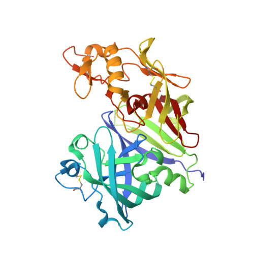
4GJ7

Crystal structure of renin in complex with NVP-BCA079 (compound 12a)


Experimental Data Snapshot

  • Method: X-RAY DIFFRACTION
  • Resolution: 2.80 Å
  • R-Value Free: 0.220 
  • R-Value Work: 0.180 
  • R-Value Observed: 0.184 

wwPDB Validation   3D Report Full Report


Ligand Structure Quality Assessment 


This is version 1.4 of the entry. See complete history


Literature

The Discovery of Novel Potent trans-3,4-Disubstituted Pyrrolidine Inhibitors of the Human Aspartic Protease Renin from in Silico Three-Dimensional (3D) Pharmacophore Searches.

Lorthiois, E.Breitenstein, W.Cumin, F.Ehrhardt, C.Francotte, E.Jacoby, E.Ostermann, N.Sellner, H.Kosaka, T.Webb, R.L.Rigel, D.F.Hassiepen, U.Richert, P.Wagner, T.Maibaum, J.

(2013) J Med Chem 56: 2207-2217

  • DOI: https://doi.org/10.1021/jm3017078
  • Primary Citation of Related Structures:  
    4GJ5, 4GJ6, 4GJ7

  • PubMed Abstract: 

    The small-molecule trans-3,4-disubstituted pyrrolidine 6 was identified from in silico three-dimensional (3D) pharmacophore searches based on known X-ray structures of renin-inhibitor complexes and demonstrated to be a weakly active inhibitor of the human enzyme. The unexpected binding mode of the more potent enantiomer (3S,4S)-6a in an extended conformation spanning the nonprime and S1' pockets of the recombinant human (rh)-renin active site was elucidated by X-ray crystallography. Initial structure-activity relationship work focused on modifications of the hydrophobic diphenylamine portion positioned in S1 and extending toward the S2 pocket. Replacement with an optimized P3-P1 pharmacophore interacting to the nonsubstrate S3(sp) cavity eventually resulted in significantly improved in vitro potency and selectivity. The prototype analogue (3S,4S)-12a of this new class of direct renin inhibitors exerted blood pressure lowering effects in a hypertensive double-transgenic rat model after oral administration.


  • Organizational Affiliation

    Novartis Pharma AG, Institutes for BioMedical Research, Novartis Campus, CH-4056 Basel, Switzerland. edwige.lorthiois@novartis.com


Macromolecules
Find similar proteins by:  (by identity cutoff)  |  3D Structure
Entity ID: 1
MoleculeChains Sequence LengthOrganismDetailsImage
Renin
A, B
340Homo sapiensMutation(s): 0 
Gene Names: REN
EC: 3.4.23.15
UniProt & NIH Common Fund Data Resources
Find proteins for P00797 (Homo sapiens)
Explore P00797 
Go to UniProtKB:  P00797
PHAROS:  P00797
GTEx:  ENSG00000143839 
Entity Groups  
Sequence Clusters30% Identity50% Identity70% Identity90% Identity95% Identity100% Identity
UniProt GroupP00797
Sequence Annotations
Expand
  • Reference Sequence
Small Molecules
Binding Affinity Annotations 
IDSourceBinding Affinity
0LT BindingDB:  4GJ7 IC50: min: 170, max: 450 (nM) from 3 assay(s)
PDBBind:  4GJ7 IC50: 170 (nM) from 1 assay(s)
Experimental Data & Validation

Experimental Data

  • Method: X-RAY DIFFRACTION
  • Resolution: 2.80 Å
  • R-Value Free: 0.220 
  • R-Value Work: 0.180 
  • R-Value Observed: 0.184 
  • Space Group: P 21 3
Unit Cell:
Length ( Å )Angle ( ˚ )
a = 141.476α = 90
b = 141.476β = 90
c = 141.476γ = 90
Software Package:
Software NamePurpose
BUSTERrefinement
DENZOdata reduction
SCALEPACKdata scaling

Structure Validation

View Full Validation Report


Ligand Structure Quality Assessment 


Entry History 

Deposition Data

Revision History  (Full details and data files)

  • Version 1.0: 2013-03-06
    Type: Initial release
  • Version 1.1: 2013-04-10
    Changes: Database references
  • Version 1.2: 2018-04-04
    Changes: Data collection
  • Version 1.3: 2020-07-29
    Type: Remediation
    Reason: Carbohydrate remediation
    Changes: Data collection, Derived calculations, Structure summary
  • Version 1.4: 2023-09-13
    Changes: Data collection, Database references, Refinement description, Structure summary