4GH6

Crystal structure of the PDE9A catalytic domain in complex with inhibitor 28


Experimental Data Snapshot

  • Method: X-RAY DIFFRACTION
  • Resolution: 2.70 Å
  • R-Value Free: 0.245 
  • R-Value Work: 0.212 

wwPDB Validation   3D Report Full Report


Ligand Structure Quality Assessment 


This is version 1.2 of the entry. See complete history


Literature

Structure-Based Discovery of Highly Selective Phosphodiesterase-9A Inhibitors and Implications for Inhibitor Design.

Meng, F.Hou, J.Shao, Y.X.Wu, P.Y.Huang, M.Zhu, X.Cai, Y.Li, Z.Xu, J.Liu, P.Luo, H.B.Wan, Y.Ke, H.

(2012) J Med Chem 55: 8549-8558

  • DOI: https://doi.org/10.1021/jm301189c
  • Primary Citation of Related Structures:  
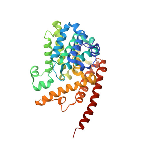
    4GH6

  • PubMed Abstract: 

    A new series of phosphodiesterase-9 (PDE9) inhibitors that contain a scaffold of 6-amino-pyrazolopyrimidinone have been discovered by a combination of structure-based design and computational docking. This procedure significantly saved the load of chemical synthesis and is an effective method for the discovery of inhibitors. The best compound 28 has an IC(50) of 21 nM and 3.3 μM, respectively, for PDE9 and PDE5 and about 3 orders of magnitude of selectivity against other PDE families. The crystal structure of the PDE9 catalytic domain in complex with 28 has been determined and shows a hydrogen bond between 28 and Tyr424. This hydrogen bond may account for the 860-fold selectivity of 28 against PDE1B, in comparison with about 30-fold selectivity of BAY73-6691. Thus, our studies suggest that Tyr424, a unique residue of PDE8 and PDE9, is a potential target for improvement of selectivity of PDE9 inhibitors.


  • Organizational Affiliation

    School of Chemistry and Chemical Engineering, Sun Yat-Sen University, Guangzhou 510275, People's Republic of China.


Macromolecules
Find similar proteins by:  (by identity cutoff)  |  3D Structure
Entity ID: 1
MoleculeChains Sequence LengthOrganismDetailsImage
High affinity cGMP-specific 3',5'-cyclic phosphodiesterase 9A
A, B
326Homo sapiensMutation(s): 0 
Gene Names: PDE9A
EC: 3.1.4.35
UniProt & NIH Common Fund Data Resources
Find proteins for O76083 (Homo sapiens)
Explore O76083 
Go to UniProtKB:  O76083
PHAROS:  O76083
GTEx:  ENSG00000160191 
Entity Groups  
Sequence Clusters30% Identity50% Identity70% Identity90% Identity95% Identity100% Identity
UniProt GroupO76083
Sequence Annotations
Expand
  • Reference Sequence
Small Molecules
Binding Affinity Annotations 
IDSourceBinding Affinity
LUO PDBBind:  4GH6 IC50: 21 (nM) from 1 assay(s)
Experimental Data & Validation

Experimental Data

  • Method: X-RAY DIFFRACTION
  • Resolution: 2.70 Å
  • R-Value Free: 0.245 
  • R-Value Work: 0.212 
  • Space Group: P 41 21 2
Unit Cell:
Length ( Å )Angle ( ˚ )
a = 104.276α = 90
b = 104.276β = 90
c = 270.073γ = 90
Software Package:
Software NamePurpose
AMoREphasing
CNSrefinement
HKL-2000data reduction
HKL-2000data scaling

Structure Validation

View Full Validation Report


Ligand Structure Quality Assessment 


Entry History 

Deposition Data

  • Released Date: 2012-10-03 
  • Deposition Author(s): Hou, J., Ke, H.

Revision History  (Full details and data files)

  • Version 1.0: 2012-10-03
    Type: Initial release
  • Version 1.1: 2012-11-14
    Changes: Database references
  • Version 1.2: 2024-02-28
    Changes: Data collection, Database references, Derived calculations