4GGZ

The structure of bradavidin2-biotin complex


Experimental Data Snapshot

  • Method: X-RAY DIFFRACTION
  • Resolution: 1.75 Å
  • R-Value Free: 0.240 
  • R-Value Work: 0.192 
  • R-Value Observed: 0.195 

wwPDB Validation   3D Report Full Report


Ligand Structure Quality Assessment 


This is version 1.2 of the entry. See complete history


Literature

The highly dynamic oligomeric structure of bradavidin II is unique among avidin proteins.

Leppiniemi, J.Meir, A.Kahkonen, N.Kukkurainen, S.Maatta, J.A.Ojanen, M.Janis, J.Kulomaa, M.S.Livnah, O.Hytonen, V.P.

(2013) Protein Sci 22: 980-994

  • DOI: https://doi.org/10.1002/pro.2281
  • Primary Citation of Related Structures:  
    4GGR, 4GGT, 4GGZ

  • PubMed Abstract: 

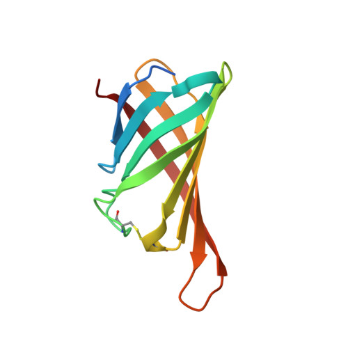
    Bradavidin II is a biotin-binding protein from Bradyrhizobium japonicum that resembles chicken avidin and bacterial streptavidin. A biophysical characterization was carried out using dynamic light scattering, native mass spectrometry, differential scanning calorimetry, and isothermal titration calorimetry combined with structural characterization using X-ray crystallography. These observations revealed that bradavidin II differs from canonical homotetrameric avidin protein family members in its quaternary structure. In contrast with the other avidins, bradavidin II appears to have a dynamic (transient) oligomeric state in solution. It is monomeric at low protein concentrations but forms higher oligomeric assemblies at higher concentrations. The crystal structure of bradavidin II revealed an important role for Phe42 in shielding the bound ligand from surrounding water molecules, thus functionally replacing the L7,8 loop essential for tight ligand binding in avidin and streptavidin. This bradavidin II characterization opens new avenues for oligomerization-independent biotin-binding protein development.


  • Organizational Affiliation

    Institute of Biomedical Technology, University of Tampere and Tampere University Hospital, FI-33014, Tampere, Finland.


Macromolecules
Find similar proteins by:  (by identity cutoff)  |  3D Structure
Entity ID: 1
MoleculeChains Sequence LengthOrganismDetailsImage
Bradavidin 2
A, B, C, D
115Bradyrhizobium diazoefficiens USDA 110Mutation(s): 0 
Gene Names: bll1558
UniProt
Find proteins for Q89U61 (Bradyrhizobium diazoefficiens (strain JCM 10833 / BCRC 13528 / IAM 13628 / NBRC 14792 / USDA 110))
Explore Q89U61 
Go to UniProtKB:  Q89U61
Entity Groups  
Sequence Clusters30% Identity50% Identity70% Identity90% Identity95% Identity100% Identity
UniProt GroupQ89U61
Sequence Annotations
Expand
  • Reference Sequence
Small Molecules
Binding Affinity Annotations 
IDSourceBinding Affinity
BTN Binding MOAD:  4GGZ Kd: 0.07 (nM) from 1 assay(s)
PDBBind:  4GGZ Kd: 0.07 (nM) from 1 assay(s)
Experimental Data & Validation

Experimental Data

  • Method: X-RAY DIFFRACTION
  • Resolution: 1.75 Å
  • R-Value Free: 0.240 
  • R-Value Work: 0.192 
  • R-Value Observed: 0.195 
  • Space Group: C 1 2 1
Unit Cell:
Length ( Å )Angle ( ˚ )
a = 119.067α = 90
b = 95.434β = 113.58
c = 49.947γ = 90
Software Package:
Software NamePurpose
HKL-2000data collection
PHASERphasing
REFMACrefinement
HKL-2000data reduction
HKL-2000data scaling

Structure Validation

View Full Validation Report


Ligand Structure Quality Assessment 


Entry History 

Deposition Data

Revision History  (Full details and data files)

  • Version 1.0: 2013-06-19
    Type: Initial release
  • Version 1.1: 2013-07-10
    Changes: Database references
  • Version 1.2: 2023-09-13
    Changes: Data collection, Database references, Derived calculations, Refinement description