4GFC

N-terminal coiled-coil dimer of C.elegans SAS-6, crystal form B


Experimental Data Snapshot

  • Method: X-RAY DIFFRACTION
  • Resolution: 2.85 Å
  • R-Value Free: 0.236 
  • R-Value Work: 0.214 
  • R-Value Observed: 0.215 

wwPDB Validation   3D Report Full Report


This is version 1.4 of the entry. See complete history


Literature

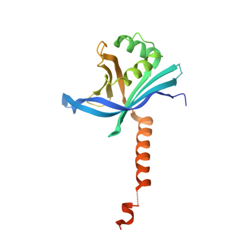
Caenorhabditis elegans centriolar protein SAS-6 forms a spiral that is consistent with imparting a ninefold symmetry.

Hilbert, M.Erat, M.C.Hachet, V.Guichard, P.Blank, I.D.Fluckiger, I.Slater, L.Lowe, E.D.Hatzopoulos, G.N.Steinmetz, M.O.Gonczy, P.Vakonakis, I.

(2013) Proc Natl Acad Sci U S A 110: 11373-11378

  • DOI: https://doi.org/10.1073/pnas.1302721110
  • Primary Citation of Related Structures:  
    4G79, 4GEU, 4GEX, 4GFA, 4GFC

  • PubMed Abstract: 

    Centrioles are evolutionary conserved organelles that give rise to cilia and flagella as well as centrosomes. Centrioles display a characteristic ninefold symmetry imposed by the spindle assembly abnormal protein 6 (SAS-6) family. SAS-6 from Chlamydomonas reinhardtii and Danio rerio was shown to form ninefold symmetric, ring-shaped oligomers in vitro that were similar to the cartwheels observed in vivo during early steps of centriole assembly in most species. Here, we report crystallographic and EM analyses showing that, instead, Caenorhabotis elegans SAS-6 self-assembles into a spiral arrangement. Remarkably, we find that this spiral arrangement is also consistent with ninefold symmetry, suggesting that two distinct SAS-6 oligomerization architectures can direct the same output symmetry. Sequence analysis suggests that SAS-6 spirals are restricted to specific nematodes. This oligomeric arrangement may provide a structural basis for the presence of a central tube instead of a cartwheel during centriole assembly in these species.


  • Organizational Affiliation

    Laboratory of Biomolecular Research (LBR), Paul Scherrer Institut, CH-5232 Villigen PSI, Switzerland.


Macromolecules
Find similar proteins by:  (by identity cutoff)  |  3D Structure
Entity ID: 1
MoleculeChains Sequence LengthOrganismDetailsImage
Spindle assembly abnormal protein 6
A, B
189Caenorhabditis elegansMutation(s): 1 
Gene Names: sas-6Y45F10D.9
UniProt
Find proteins for O62479 (Caenorhabditis elegans)
Explore O62479 
Go to UniProtKB:  O62479
Entity Groups  
Sequence Clusters30% Identity50% Identity70% Identity90% Identity95% Identity100% Identity
UniProt GroupO62479
Sequence Annotations
Expand
  • Reference Sequence
Small Molecules
Ligands 3 Unique
IDChains Name / Formula / InChI Key2D Diagram3D Interactions
PG4
Query on PG4

Download Ideal Coordinates CCD File 
I [auth A],
J [auth A]
TETRAETHYLENE GLYCOL
C8 H18 O5
UWHCKJMYHZGTIT-UHFFFAOYSA-N
IMD
Query on IMD

Download Ideal Coordinates CCD File 
G [auth A],
H [auth A]
IMIDAZOLE
C3 H5 N2
RAXXELZNTBOGNW-UHFFFAOYSA-O
EDO
Query on EDO

Download Ideal Coordinates CCD File 
C [auth A]
D [auth A]
E [auth A]
F [auth A]
K [auth A]
C [auth A],
D [auth A],
E [auth A],
F [auth A],
K [auth A],
L [auth A],
M [auth A],
N [auth B]
1,2-ETHANEDIOL
C2 H6 O2
LYCAIKOWRPUZTN-UHFFFAOYSA-N
Experimental Data & Validation

Experimental Data

  • Method: X-RAY DIFFRACTION
  • Resolution: 2.85 Å
  • R-Value Free: 0.236 
  • R-Value Work: 0.214 
  • R-Value Observed: 0.215 
  • Space Group: C 1 2 1
Unit Cell:
Length ( Å )Angle ( ˚ )
a = 103.52α = 90
b = 50.72β = 97.91
c = 101.87γ = 90
Software Package:
Software NamePurpose
HKL-2000data collection
PHASERphasing
BUSTERrefinement
MOSFLMdata reduction
SCALAdata scaling

Structure Validation

View Full Validation Report


Entry History 

Deposition Data

Revision History  (Full details and data files)

  • Version 1.0: 2013-06-19
    Type: Initial release
  • Version 1.1: 2013-07-17
    Changes: Database references
  • Version 1.2: 2013-07-24
    Changes: Database references
  • Version 1.3: 2017-08-16
    Changes: Refinement description, Source and taxonomy
  • Version 1.4: 2024-02-28
    Changes: Data collection, Database references, Derived calculations