4GD8

SHV-1 beta-lactamase in complex with penam sulfone SA3-53


Experimental Data Snapshot

  • Method: X-RAY DIFFRACTION
  • Resolution: 1.60 Å
  • R-Value Free: 0.242 
  • R-Value Work: 0.198 
  • R-Value Observed: 0.200 

wwPDB Validation   3D Report Full Report


Ligand Structure Quality Assessment 


This is version 1.1 of the entry. See complete history


Literature

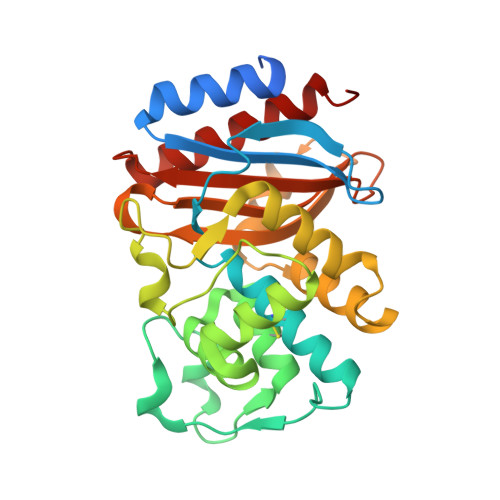
Structures of SHV-1 beta-lactamase with penem and penam sulfone inhibitors that form cyclic intermediates stabilized by carbonyl conjugation

Ke, W.Pattanaik, P.Bethel, C.R.Sheri, A.Buynak, J.D.Bonomo, R.A.van den Akker, F.

(2012) PLoS One 7: e49035-e49035

  • DOI: https://doi.org/10.1371/journal.pone.0049035
  • Primary Citation of Related Structures:  
    4GD6, 4GD8, 4GDB

  • PubMed Abstract: 

    Bacterial β-lactamase enzymes are in large part responsible for the decreased ability of β-lactam antibiotics to combat infections. The inability to overcome β-lactamase mediated resistance spurred the development of inhibitors with penems and penam sulfones being amongst the most potent and broad spectrum mechanism-based inactivators. These inhibitors form covalent, "suicide-type" inhibitory intermediates that are attached to the catalytic S70 residue. To further probe the details of the mechanism of β-lactamase inhibition by these novel compounds, we determined the crystal structures of SHV-1 bound with penem 1, and penam sulfones SA1-204 and SA3-53. Comparison with each other and with previously determined crystal structures of members of these classes of inhibitors suggests that the final conformation of the covalent adduct can vary greatly amongst the complex structures. In contrast, a common theme of carbonyl conjugation as a mechanism to avoid deacylation emerges despite that the penem and penam sulfone inhibitors form different types of intermediates. The detailed insights gained from this study could be used to further improve new mechanism-based inhibitors of these common class A serine β-lactamases.


  • Organizational Affiliation

    Department of Biochemistry, Case Western Reserve University, Cleveland, OH, USA.


Macromolecules
Find similar proteins by:  (by identity cutoff)  |  3D Structure
Entity ID: 1
MoleculeChains Sequence LengthOrganismDetailsImage
Beta-lactamase SHV-1286Klebsiella pneumoniaeMutation(s): 0 
Gene Names: blashv1
EC: 3.5.2.6
UniProt
Find proteins for P0AD64 (Klebsiella pneumoniae)
Explore P0AD64 
Go to UniProtKB:  P0AD64
Entity Groups  
Sequence Clusters30% Identity50% Identity70% Identity90% Identity95% Identity100% Identity
UniProt GroupP0AD64
Sequence Annotations
Expand
  • Reference Sequence
Small Molecules
Ligands 2 Unique
IDChains Name / Formula / InChI Key2D Diagram3D Interactions
MA4
Query on MA4

Download Ideal Coordinates CCD File 
C [auth A],
D [auth A]
CYCLOHEXYL-HEXYL-BETA-D-MALTOSIDE
C24 H44 O11
WUCWJXGMSXTDAV-QKMCSOCLSA-N
MXF
Query on MXF

Download Ideal Coordinates CCD File 
B [auth A](2S,3R)-4-(2-aminoethylcarbamoyloxy)-2-[(2-methanoylindolizin-3-yl)amino]-3-methyl-3-sulfino-butanoic acid
C17 H22 N4 O7 S
UGXGYKKZKHIUJW-GUYCJALGSA-N
Experimental Data & Validation

Experimental Data

  • Method: X-RAY DIFFRACTION
  • Resolution: 1.60 Å
  • R-Value Free: 0.242 
  • R-Value Work: 0.198 
  • R-Value Observed: 0.200 
  • Space Group: P 21 21 21
Unit Cell:
Length ( Å )Angle ( ˚ )
a = 49.394α = 90
b = 55.46β = 90
c = 83.649γ = 90
Software Package:
Software NamePurpose
Blu-Icedata collection
REFMACrefinement
d*TREKdata reduction
d*TREKdata scaling

Structure Validation

View Full Validation Report


Ligand Structure Quality Assessment 


Entry History 

Deposition Data

Revision History  (Full details and data files)

  • Version 1.0: 2013-07-31
    Type: Initial release
  • Version 1.1: 2013-08-07
    Changes: Database references