4GAB

Human AKR1B10 mutant V301L complexed with NADP+ and fidarestat


Experimental Data Snapshot

  • Method: X-RAY DIFFRACTION
  • Resolution: 1.60 Å
  • R-Value Free: 0.195 
  • R-Value Work: 0.175 
  • R-Value Observed: 0.176 

wwPDB Validation   3D Report Full Report


Ligand Structure Quality Assessment 


This is version 1.2 of the entry. See complete history


Literature

X-ray structure of the V301L aldo-keto reductase 1B10 complexed with NADP(+) and the potent aldose reductase inhibitor fidarestat: Implications for inhibitor binding and selectivity.

Ruiz, F.X.Cousido-Siah, A.Mitschler, A.Farres, J.Pares, X.Podjarny, A.

(2013) Chem Biol Interact 202: 178-185

  • DOI: https://doi.org/10.1016/j.cbi.2012.12.013
  • Primary Citation of Related Structures:  
    4GAB

  • PubMed Abstract: 

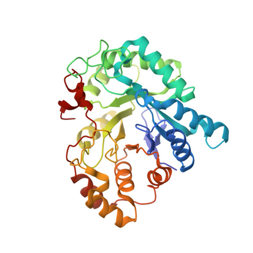
    Only one crystal structure is currently available for tumor marker AKR1B10, complexed with NADP(+) and tolrestat, which is an aldose reductase inhibitor (ARI) of the carboxylic acid type. Here, the X-ray structure of the complex of the V301L substituted AKR1B10 holoenzyme with fidarestat, an ARI of the cyclic imide type, was obtained at 1.60Å resolution by replacement soaking of crystals containing tolrestat. Previously, fidarestat was found to be safe in phase III trials for diabetic neuropathy and, consistent with its low in vivo side effects, was highly selective for aldose reductase (AR or AKR1B1) versus aldehyde reductase (AKR1A1). Now, inhibition studies showed that fidarestat was indeed 1300-fold more selective for AR as compared to AKR1B10, while the change of Val to Leu (found in AR) caused a 20-fold decrease in the IC50 value with fidarestat. Structural analysis of the V301L AKR1B10-fidarestat complex displayed enzyme-inhibitor interactions similar to those of the AR-fidarestat complex. However, a close inspection of both the new crystal structure and a computer model of the wild-type AKR1B10 complex with fidarestat revealed subtle changes that could affect fidarestat binding. In the crystal structure, a significant motion of loop A was observed between AR and V301L AKR1B10, linked to a Phe-122/Phe-123 side chain displacement. This was due to the presence of the more voluminous Gln-303 side chain (Ser-302 in AR) and of a water molecule buried in a subpocket located at the base of flexible loop A. In the wild-type AKR1B10 model, a short contact was predicted between the Val-301 side chain and fidarestat, but would not be present in AR or in V301L AKR1B10. Overall, these changes could contribute to the difference in inhibitory potency of fidarestat between AR and AKR1B10.


  • Organizational Affiliation

    Institut de Génétique et de Biologie Moléculaire et Cellulaire, Illkirch, France.


Macromolecules
Find similar proteins by:  (by identity cutoff)  |  3D Structure
Entity ID: 1
MoleculeChains Sequence LengthOrganismDetailsImage
Aldo-keto reductase family 1 member B10316Homo sapiensMutation(s): 1 
Gene Names: AKR1B10AKR1B11
EC: 1.1.1
UniProt & NIH Common Fund Data Resources
Find proteins for O60218 (Homo sapiens)
Explore O60218 
Go to UniProtKB:  O60218
PHAROS:  O60218
GTEx:  ENSG00000198074 
Entity Groups  
Sequence Clusters30% Identity50% Identity70% Identity90% Identity95% Identity100% Identity
UniProt GroupO60218
Sequence Annotations
Expand
  • Reference Sequence
Small Molecules
Ligands 3 Unique
IDChains Name / Formula / InChI Key2D Diagram3D Interactions
NAP
Query on NAP

Download Ideal Coordinates CCD File 
B [auth A]NADP NICOTINAMIDE-ADENINE-DINUCLEOTIDE PHOSPHATE
C21 H28 N7 O17 P3
XJLXINKUBYWONI-NNYOXOHSSA-N
FID
Query on FID

Download Ideal Coordinates CCD File 
C [auth A](2S,4S)-2-AMINOFORMYL-6-FLUORO-SPIRO[CHROMAN-4,4'-IMIDAZOLIDINE]-2',5'-DIONE
C12 H10 F N3 O4
WAAPEIZFCHNLKK-UFBFGSQYSA-N
CL
Query on CL

Download Ideal Coordinates CCD File 
D [auth A]CHLORIDE ION
Cl
VEXZGXHMUGYJMC-UHFFFAOYSA-M
Binding Affinity Annotations 
IDSourceBinding Affinity
FID Binding MOAD:  4GAB IC50: 3.30e+4 (nM) from 1 assay(s)
Experimental Data & Validation

Experimental Data

  • Method: X-RAY DIFFRACTION
  • Resolution: 1.60 Å
  • R-Value Free: 0.195 
  • R-Value Work: 0.175 
  • R-Value Observed: 0.176 
  • Space Group: P 61
Unit Cell:
Length ( Å )Angle ( ˚ )
a = 89.022α = 90
b = 89.022β = 90
c = 78.22γ = 120
Software Package:
Software NamePurpose
SCALEPACKdata scaling
AMoREphasing
PHENIXrefinement
PDB_EXTRACTdata extraction
HKL-2000data collection
DENZOdata reduction

Structure Validation

View Full Validation Report


Ligand Structure Quality Assessment 


Entry History 

Deposition Data

Revision History  (Full details and data files)

  • Version 1.0: 2013-03-20
    Type: Initial release
  • Version 1.1: 2013-04-10
    Changes: Database references
  • Version 1.2: 2023-09-13
    Changes: Data collection, Database references, Derived calculations, Refinement description